Application Notes: 1 µg/ml of SMC-476 was sufficient for detection of HSF1 in 20 µg of heat shocked HeLa cell lysate by colorimetric immunoblot analysis using Rabbit anti-rat IgG: AP as the secondary antibody
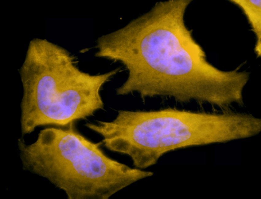
Immunofluorescence analysis of heat shocked hela cells using HSF1 antibody
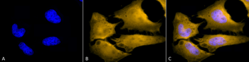
Immunofluorescence analysis of heat shocked hela cells using HSF1 antibody
Immunocytochemistry/Immunofluorescence analysis using Rat Anti-HSF1 Monoclonal Antibody, Clone 10H4. Tissue: Heat Shocked cervical cancer cells (HeLa). Species: Human. Fixation: 2% Formaldehyde for 20 min at RT. Primary Antibody: Rat Anti-HSF1 Monoclonal Antibody at 1:100 for 12 hours at 4C. Secondary Antibody: R-PE Goat Anti-Rat (yellow) at 1:200 for 2 hours at RT. Counterstain: DAPI (blue) nuclear stain at 1:40000 for 2 hours at RT. Localization: Cytoplasm. Localizes to the nucleus upon activation. Magnification: 100x. (A) DAPI (blue) nuclear stain. (B) Anti-HSF1 Antibody. (C) Composite. Heat Shocked at 42C for 1h.
Immunohistochemistry analysis using Rat Anti-HSF1 Monoclonal Antibody, Clone 10H4. Tissue: Breast carcinoma. Species: Human. Fixation: 10% Formalin Solution for 20 hours at RT. Primary Antibody: Rat Anti-HSF1 Monoclonal Antibody at 1:8000 for 40 min. Secondary Antibody: Dako labeled Polymer HRP Anti-rat IgG, DAB Chromogen (brown) (Dako Envision+ System) for 30 min at RT. Counterstain: Mayers Hematoxylin (purple/blue) nuclear stain for 1 minute at RT. Localization: Nuclear. Magnification: 100X.
Western Blot analysis of Human A431 and HEK293 cell lysates showing detection of HSF1 protein using Rat Anti-HSF1 Monoclonal Antibody, Clone 10H4. Primary Antibody: Rat Anti-HSF1 Monoclonal Antibody at 1:1000.
Immunocytochemistry/Immunofluorescence analysis using Rat Anti-HSF1 Monoclonal Antibody, Clone 10H4. Tissue: Heat Shocked cervical cancer cells (HeLa). Species: Human. Fixation: 2% Formaldehyde for 20 min at RT. Primary Antibody: Rat Anti-HSF1 Monoclonal Antibody at 1:100 for 12 hours at 4C. Secondary Antibody: FITC Goat Anti-Rat (green) at 1:200 for 2 hours at RT. Counterstain: DAPI (blue) nuclear stain at 1:40000 for 2 hours at RT. Localization: Cytoplasm. Localizes to the nucleus upon activation. Magnification: 20x. (A) DAPI (blue) nuclear stain. (B) Anti-HSF1 Antibody. (C) Composite. Heat Shocked at 42C for 1h.
Immunohistochemistry analysis using Rat Anti-HSF1 Monoclonal Antibody, Clone 10H4. Tissue: Lung. Species: Mouse. Fixation: 10% Formalin Solution for 20 hours at RT. Primary Antibody: Rat Anti-HSF1 Monoclonal Antibody at 1:1000 for 40 min. Secondary Antibody: Dako labeled Polymer HRP Anti-rat IgG, DAB Chromogen (brown) (Dako Envision+ System) for 30 min at RT. Counterstain: Mayers Hematoxylin (purple/blue) nuclear stain for 1 minute at RT. Localization: Nuclear. Magnification: 100X. (A) HSF Wildtype. (B) HSF null.
* Mehrwertsteuer und Versandkosten nicht enthalten. Irrtümer und Preisänderungen vorbehalten