E.coli-derived human CA1 recombinant protein (Position: D9-F261). Human CA1 shares 78.5% and 81% amino acid (aa) sequence identity with mouse and rat CA1, respectively.
Konjugation:
Unconjugated
Alternative Synonym:
CA I, CA1, CAB, Car1, Carbonate dehydratase I, Carbonic anhydrase 1, Carbonic anhydrase B, carbonic anhydrase I
Anti-Carbonic Anhydrase I/CA1 Antibody (monoclonal, 2B5). Tested in IHC, WB applications. This antibody reacts with Human, Mouse, Rat.
Each vial contains 4mg Trehalose, 0.9mg NaCl and 0.2mg Na2HPO4.
Formulierung:
Lyophilized
Target-Kategorie:
Carbonic anhydrase 1
Application Verdünnung:
Western blot, 0.25-0.5µg/ml, Human, Mouse, Rat Immunohistochemistry (Paraffin-embedded Section), 2-5µg/ml, Human
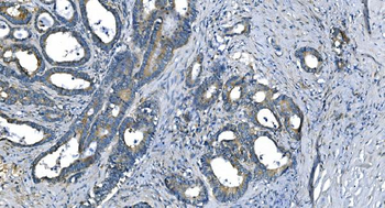
IHC analysis of Carbonic Anhydrase I/CA1 using anti-Carbonic Anhydrase I/CA1 antibody. Carbonic Anhydrase I/CA1 was detected in paraffin-embedded section of human colon cancer tissue. Heat mediated antigen retrieval was performed in EDTA buffer (pH8.0, epitope retrieval solution). The tissue section was blocked with 10% goat serum. The tissue section was then incubated with 2 µg/ml mouse anti-Carbonic Anhydrase I/CA1 Antibody overnight at 4C. Biotinylated goat anti-mouse IgG was used as secondary antibody and incubated for 30 minutes at 37C. The tissue section was developed using Strepavidin-Biotin-Complex (SABC) with DAB as the chromogen.
IHC analysis of Carbonic Anhydrase I/CA1 using anti-Carbonic Anhydrase I/CA1 antibody. Carbonic Anhydrase I/CA1 was detected in paraffin-embedded section of human gastric cancer tissue. Heat mediated antigen retrieval was performed in EDTA buffer (pH8.0, epitope retrieval solution). The tissue section was blocked with 10% goat serum. The tissue section was then incubated with 2 µg/ml mouse anti-Carbonic Anhydrase I/CA1 Antibody overnight at 4C. Biotinylated goat anti-mouse IgG was used as secondary antibody and incubated for 30 minutes at 37C. The tissue section was developed using Strepavidin-Biotin-Complex (SABC) with DAB as the chromogen.
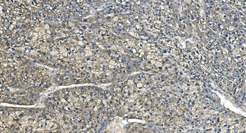
IHC analysis of Carbonic Anhydrase I/CA1 using anti-Carbonic Anhydrase I/CA1 antibody. Carbonic Anhydrase I/CA1 was detected in paraffin-embedded section of human liver cancer tissue. Heat mediated antigen retrieval was performed in EDTA buffer (pH8.0, epitope retrieval solution). The tissue section was blocked with 10% goat serum. The tissue section was then incubated with 2 µg/ml mouse anti-Carbonic Anhydrase I/CA1 Antibody overnight at 4C. Biotinylated goat anti-mouse IgG was used as secondary antibody and incubated for 30 minutes at 37C. The tissue section was developed using Strepavidin-Biotin-Complex (SABC) with DAB as the chromogen.
IHC analysis of Carbonic Anhydrase I/CA1 using anti-Carbonic Anhydrase I/CA1 antibody. Carbonic Anhydrase I/CA1 was detected in paraffin-embedded section of human lung cancer tissue. Heat mediated antigen retrieval was performed in EDTA buffer (pH8.0, epitope retrieval solution). The tissue section was blocked with 10% goat serum. The tissue section was then incubated with 2 µg/ml mouse anti-Carbonic Anhydrase I/CA1 Antibody overnight at 4C. Biotinylated goat anti-mouse IgG was used as secondary antibody and incubated for 30 minutes at 37C. The tissue section was developed using Strepavidin-Biotin-Complex (SABC) with DAB as the chromogen.
IHC analysis of Carbonic Anhydrase I/CA1 using anti-Carbonic Anhydrase I/CA1 antibody. Carbonic Anhydrase I/CA1 was detected in paraffin-embedded section of human pancreatic cancer tissue. Heat mediated antigen retrieval was performed in EDTA buffer (pH8.0, epitope retrieval solution). The tissue section was blocked with 10% goat serum. The tissue section was then incubated with 2 µg/ml mouse anti-Carbonic Anhydrase I/CA1 Antibody overnight at 4C. Biotinylated goat anti-mouse IgG was used as secondary antibody and incubated for 30 minutes at 37C. The tissue section was developed using Strepavidin-Biotin-Complex (SABC) with DAB as the chromogen.
IHC analysis of Carbonic Anhydrase I/CA1 using anti-Carbonic Anhydrase I/CA1 antibody. Carbonic Anhydrase I/CA1 was detected in paraffin-embedded section of human tonsil tissue. Heat mediated antigen retrieval was performed in EDTA buffer (pH8.0, epitope retrieval solution). The tissue section was blocked with 10% goat serum. The tissue section was then incubated with 2 µg/ml mouse anti-Carbonic Anhydrase I/CA1 Antibody overnight at 4C. Biotinylated goat anti-mouse IgG was used as secondary antibody and incubated for 30 minutes at 37C. The tissue section was developed using Strepavidin-Biotin-Complex (SABC) with DAB as the chromogen.
Western blot analysis of Carbonic Anhydrase I/CA1 using anti-Carbonic Anhydrase I/CA1 antibody. Ele
* Mehrwertsteuer und Versandkosten nicht enthalten. Irrtümer und Preisänderungen vorbehalten