
Tissue factor Recombinant Rabbit Monoclonal Antibody, Clone: [6A4], Unconjugated
Artikelnummer:
BYT-ORB704330
- Bilder (8)


| Artikelname: | Tissue factor Recombinant Rabbit Monoclonal Antibody, Clone: [6A4], Unconjugated |
| Artikelnummer: | BYT-ORB704330 |
| Hersteller Artikelnummer: | orb704330 |
| Alternativnummer: | BYT-ORB704330-50,BYT-ORB704330-100 |
| Hersteller: | Biorbyt |
| Wirt: | Rabbit |
| Kategorie: | Antikörper |
| Applikation: | ICC, IF, IHC-Fr, IHC-P, WB |
| Spezies Reaktivität: | Human, Mouse, Rat |
| Immunogen: | KLH conjugated synthetic peptide derived from human Tissue factor (1-100/295aa) |
| Konjugation: | Unconjugated |
| Alternative Synonym: | TF_HUMAN, F3, TF, Coagulation factor III, Thromboplastin, TF_MOUSE, Cf-3, Cf3, TF_RAT, |
| Tissue factor Recombinant Rabbit Monoclonal Antibody |
| Klonalität: | Recombinant |
| Konzentration: | 1mg/ml |
| Klon-Bezeichnung: | [6A4] |
| Molekulargewicht: | 35 kDa |
| UniProt: | P13726 |
| Puffer: | 0.01M TBS (pH7.4) with 1% rAlbumin, 0.02% Proclin300 and 50% Glycerol. |
| Formulierung: | Liquid |
| Target-Kategorie: | F3 |
| Application Verdünnung: | WB=1:500-2000, IHC-P=1:100-500, IHC-F=1:100-500, ICC/IF=1:50-200, IF=1:100-500 |
|
|
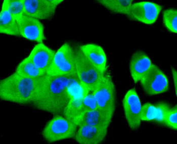
PANC-1 Hela cell, 4% Paraformaldehyde-fixed, Triton X-100 at room temperature for 20 min, Blocking buffer (normal goat serum) at 37C for 20 min, Antibody incubation with (Tissue Factor) monoclonal Antibody, Unconjugated (orb704330) 1:50, 90 minutes at 37C, followed by a conjugated Goat Anti-Rabbit IgG antibody at 37C for 90 minutes, DAPI (blue) was used to stain the cell nuclei. |
|
|
Paraformaldehyde-fixed, paraffin embedded (human breast carcinoma), Antigen retrieval by boiling in sodium citrate buffer (pH6.0) for 15 min, Block endogenous peroxidase by 3% hydrogen peroxide for 20 minutes, Blocking buffer (normal goat serum) at 37C for 30 min, Antibody incubation with (Tissue factor) Monoclonal Antibody, Unconjugated (orb704330) at 1:50 overnight at 4C, followed by operating according to SP Kit (Rabbit) instructionsand DAB staining. |
|
|
Paraformaldehyde-fixed, paraffin embedded (human pancreas tissue), Antigen retrieval by boiling in sodium citrate buffer (pH6.0) for 15 min, Block endogenous peroxidase by 3% hydrogen peroxide for 20 minutes, Blocking buffer (normal goat serum) at 37C for 30 min, Antibody incubation with (Tissue factor) Monoclonal Antibody, Unconjugated (orb704330) at 1:50 overnight at 4C, followed by operating according to SP Kit (Rabbit) instructionsand DAB staining. |
|
|
Paraformaldehyde-fixed, paraffin embedded (mouse pancreas), Antigen retrieval by boiling in sodium citrate buffer (pH6.0) for 15 min, Block endogenous peroxidase by 3% hydrogen peroxide for 20 minutes, Blocking buffer (normal goat serum) at 37C for 30 min, Antibody incubation with (Tissue factor) Monoclonal Antibody, Unconjugated (orb704330) at 1:50 overnight at 4C, followed by operating according to SP Kit (Rabbit) instructionsand DAB staining. |
|
|
Sample: Lane 1: Mouse Brain Lysates, Lane 2: Rat Kidney Lysates, Lane 3: Human A431 cell Lysates, Lane 4: Human MDA-MB-231 cell Lysates, Lane 5: Human U-87 MG cell Lysates, Lane 6: Human PANC-1 cell Lysates, Primary: Anti-Tissue factor (orb704330) at 1/1000 dilution, Secondary: IRDye800CW Goat Anti-Rabbit IgG at 1/20000 dilution, Predicted band size: 35kDa, Observed band size: 45kDa. |
|
|
Sample: Lane 1: mouse brain tissue lysate, Lane 2: U937 cell lysate, Lane 3: SH-SY5Y cell lysate, Primary: Anti-Tissue factor (orb704330) at 1:500 dilution, Secondary: Goat Anti-Rabbit IgG - HRP at 1:5000 dilution, Predicted band size: 35 kD, Observed band size: 50 kD. |
|
|
SHG-44 Hela cell, 4% Paraformaldehyde-fixed, Triton X-100 at room temperature for 20 min, Blocking buffer (normal goat serum) at 37C for 20 min, Antibody incubation with (Tissue Factor) monoclonal Antibody, Unconjugated (orb704330) 1:50, 90 minutes at 37C, followed by a conjugated Goat Anti-Rabbit IgG antibody at 37C for 90 minutes, DAPI (blue) was used to stain the cell nuclei. |
|
|
SH-SY5Y cell, 4% Paraformaldehyde-fixed, Triton X-100 at room temperature for 20 min, Blocking buffer (normal goat serum) at 37C for 20 min, Antibody incubation with (Tissue Factor) monoclonal Antibody, Unconjugated (orb704330) 1:50, 90 minutes at 37C, followed by a conjugated Goat Anti-Rabbit IgG antibody at 37C for 90 minutes, DAPI (blue) was used to stain the cell nuclei. |
Produktgarantie und fachkundiger Support
