E.coli-derived human Claudin 7/CLDN-7 recombinant protein (Position: F92-V211).
Konjugation:
Unconjugated
Alternative Synonym:
AF4/FMR2 family member 4, ALL1-fused gene from chromosome 5q31 protein, Protein AF-5q31, Major CDK9 elongation factor-associated protein, AFF4, AF5Q31, MCEF, HSPC092
Anti-Claudin 7/CLDN-7 Antibody. Tested in ELISA, Flow Cytometry, IF, IHC, ICC, WB applications. This antibody reacts with Human.
Each vial contains 4mg Trehalose, 0.9mg NaCl and 0.2mg Na2HPO4.
Formulierung:
Lyophilized
Target-Kategorie:
Claudin-7
Application Verdünnung:
Western blot, 0.1-0.25 µg/ml, Human Immunohistochemistry (Paraffin-embedded Section), 1-2µg/ml, Human Immunocytochemistry/Immunofluorescence, 5 µg/ml, Human Immunofluorescence, 5 µg/ml, Human Flow Cytometry (Fixed), 1-3µg/1x10 6 cells, Human ELISA, 0.1-0.
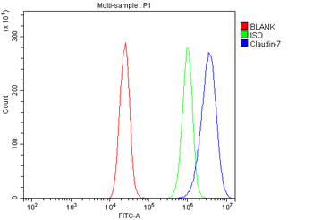
Flow Cytometry analysis of Caco-2 cells using anti-Claudin 7/CLDN-7 antibody. Overlay histogram showing Caco-2 cells (Blue line). To facilitate intracellular staining, cells were fixed with 4% paraformaldehyde and permeabilized with permeabilization buffer. The cells were blocked with 10% normal goat serum. And then incubated with rabbit anti-Claudin 7/CLDN-7 Antibody (1 µg/1x10 6 cells) for 30 min at 20C. DyLight488 conjugated goat anti-rabbit IgG (5-10 µg/1x10 6 cells) was used as secondary antibody for 30 minutes at 20C. Isotype control antibody (Green line) was rabbit IgG (1 µg/1x10 6) used under the same conditions. Unlabelled sample without incubation with primary antibody and secondary antibody (Red line) was used as a blank control.
IF analysis of Claudin 7/CLDN-7 using anti-Claudin 7/CLDN-7 antibody. Claudin 7/CLDN-7 was detected in a paraffin-embedded section of human rectal cancer tissue. Heat mediated antigen retrieval was performed in EDTA buffer (pH8.0, epitope retrieval solution). The tissue section was blocked with 10% goat serum. The tissue section was then incubated with 5 µg/mL rabbit anti-Claudin 7/CLDN-7 Antibody overnight at 4C. Biotin conjugated goat anti-rabbit IgG was used as secondary antibody and incubated for 30 minutes at 37C. The tissue section was developed using DyLight488 Conjugated Avidin. The section was counterstained with DAPI. Visualize using a fluorescence microscope and filter sets appropriate for the label used.
IF analysis of Claudin 7/CLDN-7 using anti-Claudin 7/CLDN-7 antibody. Claudin 7/CLDN-7 was detected in an immunocytochemical section of MCF-7 cells. Enzyme antigen retrieval was performed using IHC enzyme antigen retrieval reagent for 15 mins. The cells were blocked with 10% goat serum. And then incubated with 5 µg/mL rabbit anti-Claudin 7/CLDN-7 Antibody overnight at 4C. DyLight488 Conjugated Goat Anti-Rabbit IgG was used as secondary antibody at 1:100 dilution and incubated for 30 minutes at 37C. The section was counterstained with DAPI. Visualize using a fluorescence microscope and filter sets appropriate for the label used.
IHC analysis of Claudin 7/CLDN-7 using anti-Claudin 7/CLDN-7 antibody. Claudin 7/CLDN-7 was detected in a paraffin-embedded section of human gallbladder adenocarcinoma tissue. Heat mediated antigen retrieval was performed in EDTA buffer (pH8.0, epitope retrieval solution). The tissue section was blocked with 10% goat serum. The tissue section was then incubated with 2 µg/ml rabbit anti-Claudin 7/CLDN-7 Antibody overnight at 4C. Biotinylated goat anti-rabbit IgG was used as secondary antibody and incubated for 30 minutes at 37C. The tissue section was developed using Strepavidin-Biotin-Complex (SABC) with DAB as the chromogen.
IHC analysis of Claudin 7/CLDN-7 using anti-Claudin 7/CLDN-7 antibody. Claudin 7/CLDN-7 was detected in a paraffin-embedded section of human rectal cancer tissue. Heat mediated antigen retrieval was performed in EDTA buffer (pH8.0, epitope retrieval solution). The tissue section was blocked with 10% goat serum. The tissue section was then incubated with 2 µg/ml rabbit anti-Claudin 7/CLDN-7 Antibody overnight at 4C. Biotinylated goat anti-rabbit IgG was used as secondary antibody and incubated for 30 minutes at 37C. The tissue section was developed using Strepavidin-Biotin-Complex (SABC) with DAB as the chromogen.
IHC analysis of Claudin 7/CLDN-7 using anti-Claudin 7/CLDN-7 antibody. Claudin 7/CLDN-7 was detected in a paraffin-embedded section of human renal clear cell carcinoma tissue. Heat mediated antigen retrieval was performed in EDTA buffer (pH8.0, epitope retrieval solution). The tissue section was blocked with 10% goat serum. The tissue section was then incubated with 2 µg/ml rabbit anti-Claudin 7/CLDN-7 Antibody overnight at 4C. Biotinylated goat anti-rabbit IgG was used as secondary antibody and incubated for 30 minutes at 37C. The tissue section was developed using Strepavidin-Biotin-Complex (SABC) with DAB
* Mehrwertsteuer und Versandkosten nicht enthalten. Irrtümer und Preisänderungen vorbehalten