anti GJA1 antibody, anti gap junction protein, alpha 1, 43kDa antibody, anti CX43 antibody, anti DFNB38 antibody, anti GJAL antibody, anti ODDD antibody, anti connexin 43 antibody, anti gap junction 43 kDa heart protein antibody, anti gap junction protein, alpha-like antibody
Goat polyclonal antibody to GJA1
Clonality:
Polyclonal
Molecular Weight:
43
Buffer:
Supplied at 0.5 mg/ml in Tris saline, 0.02% sodium azide, pH 7.3 with 0.5% bovine serum albumin. Aliquot and store at -20C. Minimize freezing and thawing.
Sequence:
QPFDFPDDNQNSKK
Target:
Connexin 43 / GJA1
Application Dilute:
ELISA: 1:32000, WB: 0.1-1 µg/ml
Application Notes:
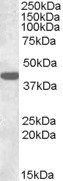
Application Notes: ELISA: Peptide ELISA: antibody detection limit dilution 1:64000.WB: Approx 40kDa band observed in Rat Brain lysates (calculated MW of 43.0kDa according to Human NP_000156.1 and to Rat NP_036699.1). Recommended concentration: 0.3-1 µg/ml
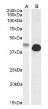
Western blot analysis of Human and Rat Heart lysate using GJA1 antibody
Western blot analysis of Human and Rat Heart lysate using GJA1 antibody
0.1 µg/mL staining of Human Cerebellum lysate (35 µg protein in RIPA buffer). Detected by chemiluminescence.
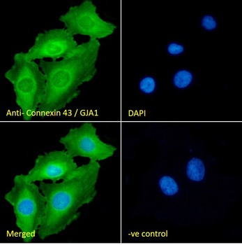
Immunofluorescence analysis of paraformaldehyde fixed HeLa cells, permeabilized with 0.15% Triton. Primary incubation 1 hr (10 µg/mL) followed by Alexa Fluor 488 secondary antibody (2 µg/mL), showing cytoplasmic and plasma membrane staining. The nuclear stain is DAPI (blue). Negative control: Unimmunized goat IgG (10 µg/mL) followed by Alexa Fluor 488 secondary antibody (2 µg/mL).
Immunofluorescence analysis of paraformaldehyde fixed U2OS cells, permeabilized with 0.15% Triton. Primary incubation 1 hr (10 µg/mL) followed by Alexa Fluor 488 secondary antibody (2 µg/mL), showing cytoplasmic, plasma membrane and nuclear staining. The nuclear stain is DAPI (blue). Negative control: Unimmunized goat IgG (10 µg/mL) followed by Alexa Fluor 488 secondary antibody (2 µg/mL).
Flow cytometric analysis of paraformaldehyde fixed HeLa cells (blue line), permeabilized with 0.5% Triton. Primary incubation 1 hr (10 µg/mL) followed by Alexa Fluor 488 secondary antibody (1 µg/mL). IgG control: Unimmunized goat IgG (black line) followed by Alexa Fluor 488 secondary antibody.
* VAT and and shipping costs not included. Errors and price changes excepted