
SPR Rabbit Polyclonal Antibody, Unconjugated
Catalog Number:
BYT-ORB158488
- Images (7)


| Article Name: | SPR Rabbit Polyclonal Antibody, Unconjugated |
| Biozol Catalog Number: | BYT-ORB158488 |
| Supplier Catalog Number: | orb158488 |
| Alternative Catalog Number: | BYT-ORB158488-50,BYT-ORB158488-100,BYT-ORB158488-200 |
| Manufacturer: | Biorbyt |
| Host: | Rabbit |
| Category: | Antikörper |
| Application: | ICC, IF, IHC-Fr, IHC-P, WB |
| Species Reactivity: | Human, Mouse, Rat |
| Immunogen: | KLH conjugated synthetic peptide derived from human Sepiapterin reductase (101-200/261aa) |
| Conjugation: | Unconjugated |
| Alternative Names: | SPRE_HUMAN, SPR, 1.1.1.153, |
| SPR Rabbit Polyclonal Antibody |
| Clonality: | Polyclonal |
| Concentration: | 1mg/ml |
| Molecular Weight: | 28 kDa |
| UniProt: | P35270 |
| Buffer: | 0.01M TBS (pH7.4) with 1% rAlbumin, 0.02% Proclin300 and 50% Glycerol. |
| Form: | Liquid |
| Target: | SPR |
| Application Dilute: | WB=1:500-2000, IHC-P=1:100-500, IHC-F=1:100-500, ICC/IF=1:100-500, IF=1:100-500 |
|
|
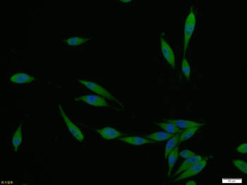
A431 cell, 4% Paraformaldehyde-fixed, Triton X-100 at room temperature for 20 min, Blocking buffer (normal goat serum) at 37C for 20 min, Antibody incubation with (SPR) polyclonal Antibody, Unconjugated (orb158488) 1:100, 90 minutes at 37C, followed by a conjugated Goat Anti-Rabbit IgG antibody at 37C for 90 minutes, DAPI (blue) was used to stain the cell nuclei. |
|
|
Paraformaldehyde-fixed, paraffin embedded (human colon carcinoma), Antigen retrieval by boiling in sodium citrate buffer (pH6.0) for 15 min, Block endogenous peroxidase by 3% hydrogen peroxide for 20 minutes, Blocking buffer (normal goat serum) at 37C for 30 min, Antibody incubation with (SPR) Polyclonal Antibody, Unconjugated (orb158488) at 1:200 overnight at 4C, followed by operating according to SP Kit (Rabbit) instructionsand DAB staining. |
|
|
Paraformaldehyde-fixed, paraffin embedded (human endometrial carcinoma), Antigen retrieval by boiling in sodium citrate buffer (pH6.0) for 15 min, Block endogenous peroxidase by 3% hydrogen peroxide for 20 minutes, Blocking buffer (normal goat serum) at 37C for 30 min, Antibody incubation with (SPR) Polyclonal Antibody, Unconjugated (orb158488) at 1:200 overnight at 4C, followed by operating according to SP Kit (Rabbit) instructionsand DAB staining. |
|
|
Paraformaldehyde-fixed, paraffin embedded (mouse pancreas), Antigen retrieval by boiling in sodium citrate buffer (pH6.0) for 15 min, Block endogenous peroxidase by 3% hydrogen peroxide for 20 minutes, Blocking buffer (normal goat serum) at 37C for 30 min, Antibody incubation with (SPR) Polyclonal Antibody, Unconjugated (orb158488) at 1:200 overnight at 4C, followed by operating according to SP Kit (Rabbit) instructionsand DAB staining. |
|
|
Paraformaldehyde-fixed, paraffin embedded (rat pancreas), Antigen retrieval by boiling in sodium citrate buffer (pH6.0) for 15 min, Block endogenous peroxidase by 3% hydrogen peroxide for 20 minutes, Blocking buffer (normal goat serum) at 37C for 30 min, Antibody incubation with (SPR) Polyclonal Antibody, Unconjugated (orb158488) at 1:200 overnight at 4C, followed by operating according to SP Kit (Rabbit) instructionsand DAB staining. |
|
|
Sample: Hela (Human) Cell Lysate at 30 ug, HepG2 (Human) Cell Lysate at 30 ug, MCF-7 (Human) Cell Lysate at 30 ug, Primary: Anti-SPR (orb158488) at 1/1000 dilution, Secondary: IRDye800CW Goat Anti-Rabbit IgG at 1/20000 dilution, Predicted band size: 28 kD, Observed band size: 28 kD. |
|
|
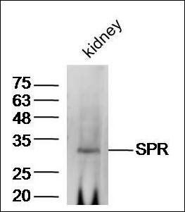
Western blot analysis of extracts from kidney using SPR antibody. (Primary dilution:1:300) |
Product Guarantee and Expert Support
