Each vial contains 4 mg Trehalose, 0.9 mg NaCl and 0.2 mg Na2HPO4.
Form:
Lyophilized
Target:
Cyclin-dependent kinase 12
Application Dilute:
Western blot, 0.25-0.5µg/ml, Human Immunohistochemistry (Paraffin-embedded Section), 2-5 µg/ml, Human Immunocytochemistry/Immunofluorescence, 5µg/ml, Human Immunoprecipitation, 0.5-2 µg/ml, Human Flow Cytometry(Fixed), 1-3µg/1x10 6 cells, Human ELISA, 0.1
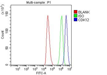
Flow Cytometry analysis of HepG2 cells using anti-CDK12 antibody. Overlay histogram showing HepG2 cells (Blue line). To facilitate intracellular staining, cells were fixed with 4% paraformaldehyde and permeabilized with permeabilization buffer. The cells were blocked with 10% normal goat serum. And then incubated with rabbit anti-CDK12 Antibody (1 µg/1x10 6 cells) for 30 min at 20C. DyLight488 conjugated goat anti-rabbit IgG (5-10 µg/1x10 6 cells) was used as secondary antibody for 30 minutes at 20C. Isotype control antibody (Green line) was rabbit IgG (1 µg/1x10 6) used under the same conditions. Unlabelled sample (Red line) was also used as a control.
IF analysis of CDK12 using anti-CDK12 antibody and anti-Beta Tubulin antibody. CDK12 was detected in immunocytochemical section of A549 cell. Enzyme antigen retrieval was performed using IHC enzyme antigen retrieval reagent for 15 mins. The cells were blocked with 10% goat serum. And then incubated with 5 µg/mL rabbit anti-CDK12 Antibody and mouse anti-Beta Tubulin antibody overnight at 4C. DyLight488 Conjugated Goat Anti-Rabbit IgG and DyLight550 Conjugated Goat Anti-Mouse IgG were used as secondary antibody at 1:500 dilution and incubated for 30 minutes at 37C. Visualize using a fluorescence microscope and filter sets appropriate for the label used.
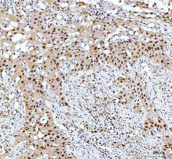
IHC analysis of CDK12 using anti-CDK12 antibody. CDK12 was detected in a paraffin-embedded section of human bladder urothelial carcinoma tissue. Heat mediated antigen retrieval was performed in EDTA buffer (pH8.0, epitope retrieval solution). The tissue section was blocked with 10% goat serum. The tissue section was then incubated with 2 µg/ml rabbit anti-CDK12 Antibody overnight at 4C. Peroxidase Conjugated Goat Anti-rabbit IgG was used as secondary antibody and incubated for 30 minutes at 37C. The tissue section was developed using HRP Conjugated Rabbit IgG Super Vision Assay Kit with DAB as the chromogen.
IHC analysis of CDK12 using anti-CDK12 antibody. CDK12 was detected in a paraffin-embedded section of human prostate cancer tissue. Heat mediated antigen retrieval was performed in EDTA buffer (pH8.0, epitope retrieval solution). The tissue section was blocked with 10% goat serum. The tissue section was then incubated with 2 µg/ml rabbit anti-CDK12 Antibody overnight at 4C. Peroxidase Conjugated Goat Anti-rabbit IgG was used as secondary antibody and incubated for 30 minutes at 37C. The tissue section was developed using HRP Conjugated Rabbit IgG Super Vision Assay Kit with DAB as the chromogen.
IHC analysis of CDK12 using anti-CDK12 antibody. CDK12 was detected in a paraffin-embedded section of human spleen tissue. Heat mediated antigen retrieval was performed in EDTA buffer (pH8.0, epitope retrieval solution). The tissue section was blocked with 10% goat serum. The tissue section was then incubated with 2 µg/ml rabbit anti-CDK12 Antibody overnight at 4C. Peroxidase Conjugated Goat Anti-rabbit IgG was used as secondary antibody and incubated for 30 minutes at 37C. The tissue section was developed using HRP Conjugated Rabbit IgG Super Vision Assay Kit with DAB as the chromogen.
Immunoprecipitating CDK12 in HepG2 whole cell lysate. Western blot analysis of CDK12 using anti-CDK12 antibody, Lane 1: HepG2 whole cell lysates (30 ug), Lane 2: Rabbit control IgG instead of anti-CDK12 antibody in HepG2 whole cell lysate, Lane 3: anti-CDK12 antibody (2 µg) + HepG2 whole cell lysate (500 µg). After electrophoresis, proteins were transferred to a membrane. Then the membrane was incubated with rabbit anti-CDK12 antigen affinity purified polyclonal antibody at a dilution of 0.5 µg/mL and probed with a goat anti-rabbit IgG-HRP secondary antibody. The signal is developed using ECL Plus Western Blotting Substrate. A specific band was detected for CDK12 at approximately 200 kDa. The expected band size for CDK12 is at 164 kDa.
Western blot analysis of CDK12 using anti-CDK12 antibody. Electrophoresis was performed on a 5-20% SDS-PA
* VAT and and shipping costs not included. Errors and price changes excepted