E.coli-derived human Desmoglein 2/DSG2 recombinant protein (Position: L24-E1020).
Alternative Synonym:
ARVC10, ARVD10, Cadherin family member 5, CDHF5, desmoglein 2, DSG2, HDGC
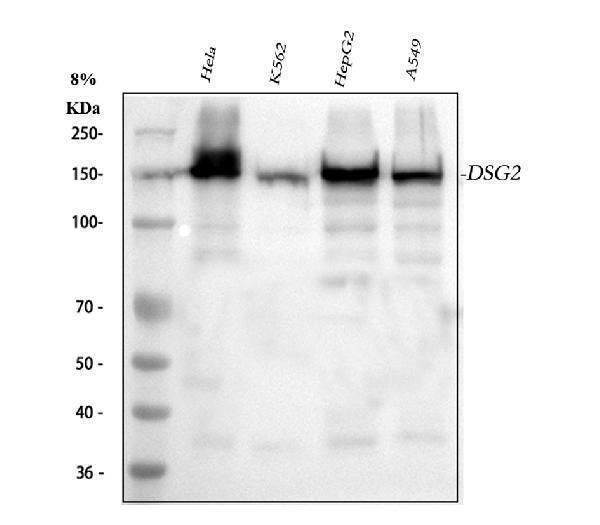
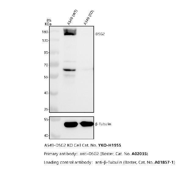
Boster Bio Anti-Desmoglein 2/DSG2 Antibody Picoband catalog A02035. Tested in ELISA, Flow Cytometry, IF, IHC, ICC, WB applications. This antibody reacts with Human. The brand Picoband indicates this is a premium antibody that guarantees superior quality, high affinity, and strong signals with minimal background in Western blot applications. Only our best-performing antibodies are designated as Picoband, ensuring unmatched performance.
Klonalität:
Polyclonal
Konzentration:
Adding 0.2 ml of distilled water will yield a concentration of 500 µg/ml.
Western blot, 0.25-0.5µg/ml, Human Immunohistochemistry (Paraffin-embedded Section), 0.5-1µg/ml, Human Immunocytochemistry/Immunofluorescence, 2µg/ml, Human Flow Cytometry (Fixed), 1-3µg/1x106 cells, Human ELISA, 0.1-0.5µg/ml, -
Flow Cytometry analysis of U87 cells using anti-DSG2 antibody (A02035). Overlay histogram showing U87 cells stained with A02035 (Blue line). To facilitate intracellular staining, cells were fixed with 4% paraformaldehyde and permeabilized with permeabilization buffer. The cells were blocked with 10% normal goat serum. And then incubated with rabbit anti-DSG2 Antibody (A02035&44, 1µg/1x106 cells) for 30 min at 20C. DyLight488 conjugated goat anti-rabbit IgG (BA1127&44, 5-10µg/1x106 cells) was used as secondary antibody for 30 minutes at 20C. Isotype control antibody (Green line) was rabbit IgG (1µg/1x106) used under the same conditions. Unlabelled sample without incubation with primary antibody and secondary antibody (Red line) was used as a blank control.
IHC analysis of DSG2 using anti-DSG2 antibody (A02035). DSG2 was detected in paraffin-embedded section of human appendicitis tissue. Heat mediated antigen retrieval was performed in EDTA buffer (pH8.0&44, epitope retrieval solution). The tissue section was blocked with 10% goat serum. The tissue section was then incubated with 1µg/ml rabbit anti-DSG2 Antibody (A02035) overnight at 4C. Biotinylated goat anti-rabbit IgG was used as secondary antibody and incubated for 30 minutes at 37C. The tissue section was developed using Strepavidin-Biotin-Complex (SABC) (Catalog SA1022) with DAB as the chromogen.
IHC analysis of DSG2 using anti-DSG2 antibody (A02035). DSG2 was detected in paraffin-embedded section of human prostatic cancer tissue. Heat mediated antigen retrieval was performed in EDTA buffer (pH8.0&44, epitope retrieval solution). The tissue section was blocked with 10% goat serum. The tissue section was then incubated with 1µg/ml rabbit anti-DSG2 Antibody (A02035) overnight at 4C. Biotinylated goat anti-rabbit IgG was used as secondary antibody and incubated for 30 minutes at 37C. The tissue section was developed using Strepavidin-Biotin-Complex (SABC) (Catalog SA1022) with DAB as the chromogen.
IHC analysis of DSG2 using anti-DSG2 antibody (A02035). DSG2 was detected in paraffin-embedded section of human rectal cancer tissue. Heat mediated antigen retrieval was performed in EDTA buffer (pH8.0&44, epitope retrieval solution). The tissue section was blocked with 10% goat serum. The tissue section was then incubated with 1µg/ml rabbit anti-DSG2 Antibody (A02035) overnight at 4C. Biotinylated goat anti-rabbit IgG was used as secondary antibody and incubated for 30 minutes at 37C. The tissue section was developed using Strepavidin-Biotin-Complex (SABC) (Catalog SA1022) with DAB as the chromogen.
Flow Cytometry analysis of HEPA 1-6 cells using anti-DSG2 antibody (A02035). Overlay histogram showing HEPA 1-6 cells stained with A02035 (Blue line). To facilitate intracellular staining, cells were fixed with 4% paraformaldehyde and permeabilized with permeabilization buffer. The cells were blocked with 10% normal goat serum. And then incubated with rabbit anti-DSG2 Antibody (A02035&44, 1µg/1x106 cells) for 30 min at 20C. DyLight488 conjugated goat anti-rabbit IgG (BA1127&44, 5-10µg/1x10
Flow Cytometry analysis of RH35 cells using anti-DSG2 antibody (A02035). Overlay histogram showing RH35 cells stained with A02035 (Blue line). To facilitate intracellular staining, cells were fixed with 4% paraformaldehyde and permeabilized with permeabilization buffer. The cells were blocked with 10% normal goat serum. And then incubated with rabbit anti-DSG2 Antibody (A02035&44, 1µg/1x106 cells) for 30 min at 20C. DyLight488 conjugated goat anti-rabbit IgG (BA1127&44, 5-10µg/1x106 cells) was used as secondary antibody for 30 minutes at 20C. Isotype control antibody (Green line) was rabbit IgG (1µg/1x106) used under the same conditions. Unlabelled sample without incubation with primary antibody and secondary antibody (Red line) was used as a blank control.
* Mehrwertsteuer und Versandkosten nicht enthalten. Irrtümer und Preisänderungen vorbehalten