
CD46 Rabbit Polyclonal Antibody, Unconjugated
Artikelnummer:
BYT-ORB10329
- Bilder (6)


| Artikelname: | CD46 Rabbit Polyclonal Antibody, Unconjugated |
| Artikelnummer: | BYT-ORB10329 |
| Hersteller Artikelnummer: | orb10329 |
| Alternativnummer: | BYT-ORB10329-50,BYT-ORB10329-100,BYT-ORB10329-200 |
| Hersteller: | Biorbyt |
| Wirt: | Rabbit |
| Kategorie: | Antikörper |
| Applikation: | FC, IF, IHC-Fr, IHC-P, WB |
| Spezies Reaktivität: | Human, Mouse |
| Immunogen: | KLH conjugated synthetic peptide derived from human CD46 (251-355/355aa) |
| Konjugation: | Unconjugated |
| Alternative Synonym: | Mcp, AHUS2, MIC10, TLX, TRA2.10, MCP_HUMAN, CD46, Trophoblast leukocyte common antigen, MCP_MOUSE, MCP_RAT, |
| CD46 Rabbit Polyclonal Antibody |
| Klonalität: | Polyclonal |
| Konzentration: | 1mg/ml |
| Molekulargewicht: | 43 kDa |
| UniProt: | P15529 |
| Puffer: | 0.01M TBS (pH7.4) with 1% rAlbumin, 0.02% Proclin300 and 50% Glycerol. |
| Formulierung: | Liquid |
| Target-Kategorie: | CD46 |
| Application Verdünnung: | WB=1:500-2000, IHC-P=1:100-500, IHC-F=1:100-500, IF=1:100-500, Flow-Cyt=1µg /test |
|
|
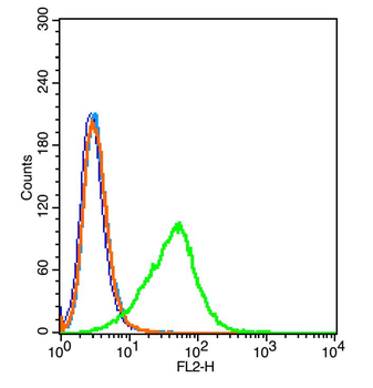
Blank control: 293T (blue). Primary Antibody: Rabbit Anti-CD46 antibody (orb10329), Dilution: 1 µg in 100 1 µl 1X PBS containing 0.5% BSA (green), Isotype Control Antibody: Rabbit IgG (orange), used under the same conditions, Secondary Antibody: Goat anti-rabbit IgG-PE (white blue), Dilution: 1:200 in 1 X PBS containing 0.5% BSA. protocol, The cells were washed twice with phosphate-buffered saline (PBS). The cells were then incubated in 1 X PBS containing 0.5% BSA + 10% goat serum (15 min) to block non-specific protein-protein interactions followed by the antibody (orb10329, 1 µg/1x10 6 cells) for 30 min on ice. The secondary antibody used was Goat Anti-rabbit IgG/PE antibody at 1/200 dilution for 30 min on ice. Acquisition of 20000 events was performed. |
|
|
Blank control: U937 (blue). Primary Antibody: Rabbit Anti-CD46 antibody (orb10329), Dilution: 1 µg in 100 µL 1X PBS containing 0.5% BSA, Isotype Control Antibody: Rabbit IgG (orange), used under the same conditions, Secondary Antibody: Goat anti-rabbit IgG-PE (white blue), Dilution: 1:200 in 1 X PBS containing 0.5% BSA. Protocol, The cells were fixed with 2% paraformaldehyde (10 min). Primary antibody (orb10329, 1 µg/1x10 6 cells) were incubated for 30 min on the ice, followed by 1 X PBS containing 0.5% BSA + 1 0% goat serum (15 min) to block non-specific protein-protein interactions. Then the Goat Anti-rabbit IgG/PE antibody was added into the blocking buffer mentioned above to react with the primary antibody at 1/200 dilution for 30 min on ice. Acquisition of 20000 events was performed. |
|
|
Paraformaldehyde-fixed, paraffin embedded (Human colon carcinoma), Antigen retrieval by boiling in sodium citrate buffer (pH6.0) for 15 min, Block endogenous peroxidase by 3% hydrogen peroxide for 20 minutes, Blocking buffer (normal goat serum) at 37C for 30 min, Antibody incubation with (CD46) Polyclonal Antibody, Unconjugated (orb10329) at 1:400 overnight at 4C, followed by operating according to SP Kit (Rabbit) instructionsand DAB staining. |
|
|
Sample: Epididymis (Mouse) Lysate at 40 ug, Primary: Anti-CD46 (orb10329) at 1/1000 dilution, Secondary: IRDye800CW Goat Anti-Rabbit IgG at 1/20000 dilution, Predicted band size: 43 kD, Observed band size: 56 kD. |
|
|
Sample: Lane 1: Mouse Placenta tissue lysates, Lane 2: Mouse Lung tissue lysates, Lane 3: Human K562 cell lysates, Lane 4: Human HeLa cell lysates, Lane 5: Human MOLT4 cell lysates, Lane 6: Human MCF-7 cell lysates, Lane 7: Human Daudi cell lysates, Primary: Anti-CD46 (orb10329) at 1/1000 dilution, Secondary: IRDye800CW Goat Anti-Rabbit IgG at 1/20000 dilution, Predicted band size: 43 kDa, Observed band size: 52 kDa. |
|
|
Sample: Testis (Mouse) Lysate at 40 ug, Primary: Anti-CD46 (orb10329) at 1/1000 dilution, Secondary: IRDye800CW Goat Anti-Rabbit IgG at 1/20000 dilution, Predicted band size: 43 kD, Observed band size: 55 kD. |
Produktgarantie und fachkundiger Support
