E.coli-derived human Sigma1-receptor/SIGMAR1 recombinant protein (Position: G1-H154).
Konjugation:
Unconjugated
Alternative Synonym:
Heat shock protein beta-8, HspB8, Alpha-crystallin C chain, E2-induced gene 1 protein, Protein kinase H11, Small stress protein-like protein HSP22, HSPB8, CRYAC, E2IG1, HSP22, PP1629
Anti-Sigma1-receptor/SIGMAR1 Antibody. Tested in ELISA, IHC, WB applications. This antibody reacts with Human, Monkey, Mouse, Rat.
Klonalität:
Polyclonal
Konzentration:
Adding 0.2 ml of distilled water will yield a concentration of 500 µg/ml.
Western blot, 0.1-0.25 µg/ml, Human, Monkey, Mouse Immunohistochemistry(Paraffin-embedded Section), 2-5 µg/ml, Human, Mouse, Rat ELISA, 0.1-0.5 µg/ml, -
IHC analysis of Sigma1-Receptor/SIGMAR1 using anti-Sigma1-Receptor/SIGMAR1 antibody. Sigma1-Receptor/SIGMAR1 was detected in a paraffin-embedded section of human adenocarcinoma of the right colon tissue was blocked with 10% goat serum. The tissue section was then incubated with 2 µg/ml rabbit anti-Sigma1-Receptor/SIGMAR1 Antibody overnight at 4C. Peroxidase Conjugated Goat Anti-rabbit IgG was used as secondary antibody and incubated for 30 minutes at 37C. The tissue section was developed using HRP Conjugated Rabbit IgG Super Vision Assay Kit with DAB as the chromogen.
IHC analysis of Sigma1-Receptor/SIGMAR1 using anti-Sigma1-Receptor/SIGMAR1 antibody. Sigma1-Receptor/SIGMAR1 was detected in a paraffin-embedded section of human liver cancer tissue was blocked with 10% goat serum. The tissue section was then incubated with 2 µg/ml rabbit anti-Sigma1-Receptor/SIGMAR1 Antibody overnight at 4C. Peroxidase Conjugated Goat Anti-rabbit IgG was used as secondary antibody and incubated for 30 minutes at 37C. The tissue section was developed using HRP Conjugated Rabbit IgG Super Vision Assay Kit with DAB as the chromogen.
IHC analysis of Sigma1-Receptor/SIGMAR1 using anti-Sigma1-Receptor/SIGMAR1 antibody. Sigma1-Receptor/SIGMAR1 was detected in a paraffin-embedded section of human stomach cancer tissue was blocked with 10% goat serum. The tissue section was then incubated with 2 µg/ml rabbit anti-Sigma1-Receptor/SIGMAR1 Antibody overnight at 4C. Peroxidase Conjugated Goat Anti-rabbit IgG was used as secondary antibody and incubated for 30 minutes at 37C. The tissue section was developed using HRP Conjugated Rabbit IgG Super Vision Assay Kit with DAB as the chromogen.
IHC analysis of Sigma1-Receptor/SIGMAR1 using anti-Sigma1-Receptor/SIGMAR1 antibody. Sigma1-Receptor/SIGMAR1 was detected in a paraffin-embedded section of mouse colon tissue. Heat mediated antigen retrieval was performed in EDTA buffer (pH8.0, epitope retrieval solution). The tissue section was blocked with 10% goat serum. The tissue section was then incubated with 2 µg/ml rabbit anti-Sigma1-Receptor/SIGMAR1 Antibody overnight at 4C. Peroxidase Conjugated Goat Anti-rabbit IgG was used as secondary antibody and incubated for 30 minutes at 37C. The tissue section was developed using HRP Conjugated Rabbit IgG Super Vision Assay Kit with DAB as the chromogen.
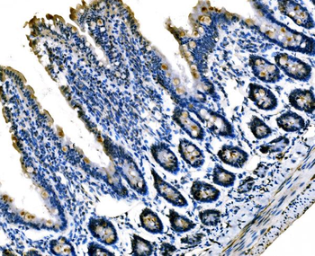
IHC analysis of Sigma1-Receptor/SIGMAR1 using anti-Sigma1-Receptor/SIGMAR1 antibody. Sigma1-Receptor/SIGMAR1 was detected in a paraffin-embedded section of rat colon tissue. Heat mediated antigen retrieval was performed in EDTA buffer (pH8.0, epitope retrieval solution). The tissue section was blocked with 10% goat serum. The tissue section was then incubated with 2 µg/ml rabbit anti-Sigma1-Receptor/SIGMAR1 Antibody overnight at 4C. Peroxidase Conjugated Goat Anti-rabbit IgG was used as secondary antibody and incubated for 30 minutes at 37C. The tissue section was developed using HRP Conjugated Rabbit IgG Super Vision Assay Kit with DAB as the chromogen.
Western blot analysis of Sigma1-Receptor/SIGMAR1 using anti-Sigma1-Receptor/SIGMAR1 antibody. Electrophoresis was performed on a 5-20% SDS-PAGE gel at 70V (Stacking gel) / 90V (Resolving gel) for 2-3 hours. The sample well of each lane was loaded with 30 ug of sample under reducing conditions. Lane 1: human Hela whole cell lysates, Lane 2: human Caco-2 whole cell lysates, Lane 3: human 293T whole cell lysates, Lane 4: human HepG2 whole cell lysates, Lane 5: human A549 whole cell lysates, Lane 6: human COLO-320 whole cell lysates, Lane 7: human U-87MG whole cell lysates, Lane 8: monkey COS-7 whole cell lysates, Lane 9: mouse liver tissue lysates, Lane 10: mouse C2C12 whole cell lysates. After electrophoresis, proteins were transferred to a nitrocellulose membrane at 150 mA for 50-90 minutes. Blocked the membrane with 5% non-fat milk/TBS for 1.5 hour at RT. The membrane was incubated with rabbit anti-Sigma1-Receptor/SIGMAR1 antigen affinity purified polyclonal ant
IHC analysis of Sigma1-Receptor/SIGMAR1 using anti-Sigma1-Receptor/SIGMAR1 antib
* Mehrwertsteuer und Versandkosten nicht enthalten. Irrtümer und Preisänderungen vorbehalten