A synthetic peptide corresponding to a sequence in the middle region of human SPG3A/ATL1, which shares 93.8% and 100% amino acid (aa) sequence identity with mouse and rat ATL1, respectively.
Konjugation:
Unconjugated
Alternative Synonym:
GBP3, SPG3A, ATL1, GBP-3, Atlastin-1, hGBP3
Anti-SPG3A/ATL1 Antibody. Tested in Flow Cytometry, IHC, WB applications. This antibody reacts with Human.
Klonalität:
Polyclonal
Konzentration:
Adding 0.2 ml of distilled water will yield a concentration of 500 µg/ml.
Western blot, 0.25-0.5 µg/ml, Human Immunohistochemistry(Paraffin-embedded Section), 2-5 µg/ml, Human Flow Cytometry (Fixed), 1-3 µg/1x10 6 cells, Human
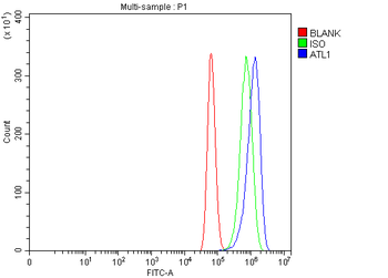
Flow Cytometry analysis of U87 cells using anti-SPG3A/ATL1 antibody. Overlay histogram showing U87 cells (Blue line). To facilitate intracellular staining, cells were fixed with 4% paraformaldehyde and permeabilized with permeabilization buffer. The cells were blocked with 10% normal goat serum. And then incubated with rabbit anti-SPG3A/ATL1 Antibody (1 µg/1x10 6 cells) for 30 min at 20C. DyLight488 conjugated goat anti-rabbit IgG (5-10 µg/1x10 6 cells) was used as secondary antibody for 30 minutes at 20C. Isotype control antibody (Green line) was rabbit IgG (1 µg/1x10 6) used under the same conditions. Unlabelled sample without incubation with primary antibody and secondary antibody (Red line) was used as a blank control.
IHC analysis of SPG3A/ATL1 using anti-SPG3A/ATL1 antibody. SPG3A/ATL1 was detected in a paraffin-embedded section of human adenocarcinoma of the right colon tissue. Heat mediated antigen retrieval was performed in EDTA buffer (pH8.0, epitope retrieval solution). The tissue section was blocked with 10% goat serum. The tissue section was then incubated with 2 µg/ml rabbit anti-SPG3A/ATL1 Antibody overnight at 4C. Peroxidase Conjugated Goat Anti-rabbit IgG was used as secondary antibody and incubated for 30 minutes at 37C. The tissue section was developed using HRP Conjugated Rabbit IgG Super Vision Assay Kit with DAB as the chromogen.
IHC analysis of SPG3A/ATL1 using anti-SPG3A/ATL1 antibody. SPG3A/ATL1 was detected in a paraffin-embedded section of human cervical cancer tissue. Heat mediated antigen retrieval was performed in EDTA buffer (pH8.0, epitope retrieval solution). The tissue section was blocked with 10% goat serum. The tissue section was then incubated with 2 µg/ml rabbit anti-SPG3A/ATL1 Antibody overnight at 4C. Peroxidase Conjugated Goat Anti-rabbit IgG was used as secondary antibody and incubated for 30 minutes at 37C. The tissue section was developed using HRP Conjugated Rabbit IgG Super Vision Assay Kit with DAB as the chromogen.
IHC analysis of SPG3A/ATL1 using anti-SPG3A/ATL1 antibody. SPG3A/ATL1 was detected in a paraffin-embedded section of human lymphoma tissue. Heat mediated antigen retrieval was performed in EDTA buffer (pH8.0, epitope retrieval solution). The tissue section was blocked with 10% goat serum. The tissue section was then incubated with 2 µg/ml rabbit anti-SPG3A/ATL1 Antibody overnight at 4C. Peroxidase Conjugated Goat Anti-rabbit IgG was used as secondary antibody and incubated for 30 minutes at 37C. The tissue section was developed using HRP Conjugated Rabbit IgG Super Vision Assay Kit with DAB as the chromogen.
IHC analysis of SPG3A/ATL1 using anti-SPG3A/ATL1 antibody. SPG3A/ATL1 was detected in a paraffin-embedded section of human stomach cancer tissue. Heat mediated antigen retrieval was performed in EDTA buffer (pH8.0, epitope retrieval solution). The tissue section was blocked with 10% goat serum. The tissue section was then incubated with 2 µg/ml rabbit anti-SPG3A/ATL1 Antibody overnight at 4C. Peroxidase Conjugated Goat Anti-rabbit IgG was used as secondary antibody and incubated for 30 minutes at 37C. The tissue section was developed using HRP Conjugated Rabbit IgG Super Vision Assay Kit with DAB as the chromogen.
Western blot analysis of SPG3A/ATL1 using anti-SPG3A/ATL1 antibody. Electrophoresis was performed on a 5-20% SDS-PAGE gel at 70V (Stacking gel) / 90V (Resolving gel) for 2-3 hours. The sample well of each lane was loaded with 30 ug of sample under reducing conditions. Lane 1: human 293T whole cell lysates, Lane 2: human SiHa whole cell lysates, Lane 3: human SH-SY5Y whole cell lysates. After electrophoresis, proteins were transferred to a nitrocellulose membrane at 150 mA for 50-90 minutes. Blocked the membrane with 5% non-fat milk/TBS for 1.5 hour at RT. The membrane was incubated with rabbit anti-SPG3A/ATL1 antigen affinity purified polyclonal antibody at 0.5 µg/mL overnight at 4C, then washed with TBS-0.1% Tween 3 times with 5 minutes each
IHC analysis of SPG3A/ATL1 using anti-SPG3A/ATL1 antibody. SPG3A/ATL1 was detected in a p
* Mehrwertsteuer und Versandkosten nicht enthalten. Irrtümer und Preisänderungen vorbehalten