
PIK3R1 Rabbit Polyclonal Antibody, Unconjugated
Artikelnummer:
BYT-ORB11271
- Bilder (7)


| Artikelname: | PIK3R1 Rabbit Polyclonal Antibody, Unconjugated |
| Artikelnummer: | BYT-ORB11271 |
| Hersteller Artikelnummer: | orb11271 |
| Alternativnummer: | BYT-ORB11271-50,BYT-ORB11271-100,BYT-ORB11271-200 |
| Hersteller: | Biorbyt |
| Wirt: | Rabbit |
| Kategorie: | Antikörper |
| Applikation: | FC, ICC, IF, IHC-Fr, IHC-P, WB |
| Spezies Reaktivität: | Human, Mouse, Rat |
| Immunogen: | KLH conjugated synthetic peptide derived from human PI3 kinase p85 subunit alpha (501-600/724aa) |
| Konjugation: | Unconjugated |
| Alternative Synonym: | AGM7, GRB1, IMD36, p85, p85-ALPHA, p85alpha, PI3K, p50alpha, p55alpha, PI3KA, P85A_HUMAN, PIK3R1, PI3-kinase regulatory subunit alpha, PI3K regulatory subunit alpha, PtdIns-3-kinase regulatory subunit alpha, Phosphatidylinositol 3-kinase 85 kDa regulatory subunit alpha (PI3-kinase subunit p85-alpha | PtdIns-3-kinase regulatory subunit p85-alpha), P85A_MOUSE, P85A_RAT, |
| PIK3R1 Rabbit Polyclonal Antibody |
| Application Verdünnung: | WB=1:500-2000, IHC-P=1:100-500, IHC-F=1:100-500, ICC/IF=1:100-500, IF=1:100-500, Flow-Cyt=1µg/Test |
|
|
Paraformaldehyde-fixed, paraffin embedded (mouse skeletal muscle), Antigen retrieval by boiling in sodium citrate buffer (pH6.0) for 15 min, Block endogenous peroxidase by 3% hydrogen peroxide for 20 minutes, Blocking buffer (normal goat serum) at 37C for 30 min, Antibody incubation with (PI 3 Kinase p85 alpha) Polyclonal Antibody, Unconjugated (orb11271) at 1:200 overnight at 4C, followed by operating according to SP Kit (Rabbit) instructionsand DAB staining. |
|
|
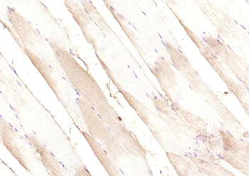
Paraformaldehyde-fixed, paraffin embedded (rat skeletal muscle), Antigen retrieval by boiling in sodium citrate buffer (pH6.0) for 15 min, Block endogenous peroxidase by 3% hydrogen peroxide for 20 minutes, Blocking buffer (normal goat serum) at 37C for 30 min, Antibody incubation with (PI 3 Kinase p85 alpha) Polyclonal Antibody, Unconjugated (orb11271) at 1:200 overnight at 4C, followed by operating according to SP Kit (Rabbit) instructionsand DAB staining. |
|
|
Positive control: H9C2 (2% Paraformaldehyde-fixed), Isotype Control Antibody, Antibody: Rabbit IgG, Dilution: 1 µg in 100 µl 1 X PBS containing 0.5% BSA, Secondary Antibody, Antibody: Goat anti-rabbit IgG-FITC, Dilution: 1:200 in 1 X PBS containing 0.5% BSA, Primary Antibody, orb10295, Dilution: 1 µg in 100 µl 1X PBS containing 0.5% BSA. |
|
|
Sample: Heart (mouse) Lysate at 40 ug, Primary: Anti-PI3K p85 (orb11271) at 1/300 dilution, Secondary: IRDye800CW Goat Anti-Rabbit IgG at 1/20000 dilution, Predicted band size: 80kD, Observed band size: 85 kD. |
|
|
Tissue/Cell: HepG2 cell, 4% Paraformaldehyde-fixed, Triton X-100 at room temperature for 20 min, Blocking buffer (normal goat serum) at 37C for 20 min, Antibody incubation with (PI3K p85) polyclonal Antibody, Unconjugated (orb11271) 1:100, 90 minutes at 37C, followed by a FITC conjugated Goat Anti-Rabbit IgG antibody at 37C for 90 minutes, DAPI (blue) was used to stain the cell nuclei. |
|
|
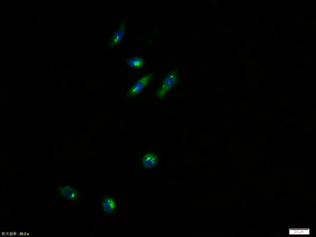
Tissue/Cell: NIH/3T3 cell, 4% Paraformaldehyde-fixed, Triton X-100 at room temperature for 20 min, Blocking buffer (normal goat serum) at 37C for 20 min, Antibody incubation with (PI 3 Kinase p85 alpha) polyclonal Antibody, Unconjugated (orb11271) 1:100, 90 minutes at 37C, followed by a FITC conjugated Goat Anti-Rabbit IgG antibody at 37C for 90 minutes, DAPI (blue) was used to stain the cell nuclei. |
|
|
Tissue/Cell: rat brain tissue, 4% Paraformaldehyde-fixed and paraffin-embedded, Antigen retrieval: citrate buffer (0.01M, pH6.0), Boiling bathing for 15 min, Block endogenous peroxidase by 3% Hydrogen peroxide for 30 min, Blocking buffer (normal goat serum) at 37C for 20 min, Incubation: Anti-PI3K/PI3 kinase p85 alpha subunit Polyclonal Antibody, Unconjugated (orb11271) 1:200, overnight at 4C, followed by conjugation to the secondary antibody and DAB staining. |
Produktgarantie und fachkundiger Support
