
TrkA Rabbit Polyclonal Antibody, Unconjugated
Artikelnummer:
BYT-ORB11515
- Bilder (6)


| Artikelname: | TrkA Rabbit Polyclonal Antibody, Unconjugated |
| Artikelnummer: | BYT-ORB11515 |
| Hersteller Artikelnummer: | orb11515 |
| Alternativnummer: | BYT-ORB11515-50,BYT-ORB11515-100,BYT-ORB11515-200 |
| Hersteller: | Biorbyt |
| Wirt: | Rabbit |
| Kategorie: | Antikörper |
| Applikation: | IF, IHC-Fr, IHC-P, WB |
| Spezies Reaktivität: | Human, Mouse, Rat |
| Immunogen: | KLH conjugated synthetic peptide derived from human Trk A (725-796/796aa) |
| Konjugation: | Unconjugated |
| Alternative Synonym: | MTC, TRK, TRK1, TRKA, Trk-A, p140-TrkA, Tkr, NTRK1_HUMAN, NTRK1, Neurotrophic tyrosine kinase receptor type 1, TRK1-transforming tyrosine kinase protein, Tropomyosin-related kinase A, Tyrosine kinase receptor, Tyrosine kinase receptor A (Trk-A), gp140trk, 2.7.10.1, NTRK1_MOUSE, NTRK1_RAT, Slow nerve growth factor receptor, p140-TrkA (Trk-A), |
| TrkA Rabbit Polyclonal Antibody |
| Klonalität: | Polyclonal |
| Konzentration: | 1mg/ml |
| Molekulargewicht: | 90 kDa |
| UniProt: | P04629 |
| Puffer: | 0.01M TBS (pH7.4) with 1% rAlbumin, 0.02% Proclin300 and 50% Glycerol. |
| Formulierung: | Liquid |
| Target-Kategorie: | NTRK1 |
| Application Verdünnung: | WB=1:500-2000, IHC-P=1:100-500, IHC-F=1:100-500, IF=1:100-500 |
|
|
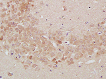
Paraformaldehyde-fixed, paraffin embedded (Mouse brain), Antigen retrieval by boiling in sodium citrate buffer (pH6.0) for 15 min, Block endogenous peroxidase by 3% hydrogen peroxide for 20 minutes, Blocking buffer (normal goat serum) at 37C for 30 min, Antibody incubation with (TrkA) Polyclonal Antibody, Unconjugated (orb11515) at 1:400 overnight at 4C, followed by operating according to SP Kit (Rabbit) instructionsand DAB staining. |
|
|
Sample: Lane 1: Cerebrum (Mouse) Lysate at 40 ug, Primary: Anti-TrkA (orb11515) at 1/1000 dilution, Secondary: IRDye800CW Goat Anti-Rabbit IgG at 1/20000 dilution, Predicted band size: 130 kD, Observed band size: 130 kD. |
|
|
Sample: Lymph node (Mouse) Lysate at 40 ug, Lymph node (Rat) Lysate at 40 ug, Primary: Anti-TrkA (orb11515) at 1/300 dilution, Secondary: IRDye800CW Goat Anti-Rabbit IgG at 1/20000 dilution, Predicted band size: 90 kD, Observed band size: 100 kD. |
|
|
Sample: U251 Cell (Human) Lysate at 40 ug, Primary: Anti-TrkA (orb11515) at 1/300 dilution, Secondary: IRDye800CW Goat Anti-Rabbit IgG at 1/20000 dilution, Predicted band size: 90 kD, Observed band size: 100 kD. |
|
|
Tissue/Cell: mouse brain tissue, 4% Paraformaldehyde-fixed and paraffin-embedded, Antigen retrieval: citrate buffer (0.01M, pH6.0), Boiling bathing for 15 min, Blocking buffer (normal goat serum) at 37C for 20 min, Incubation: Anti-TrkA Polyclonal Antibody, Unconjugated (orb11515) 1:200, overnight at 4C, The secondary antibody was Goat Anti-Rabbit IgG, Cy3 conjugated (orb868589) used at 1:200 dilution for 40 minutes at 37C. DAPI (5 ug/ml, blue) was used to stain the cell nuclei. |
|
|
Tissue/Cell: mouse embryo tissue, 4% Paraformaldehyde-fixed and paraffin-embedded, Antigen retrieval: citrate buffer (0.01M, pH6.0), Boiling bathing for 15 min, Blocking buffer (normal goat serum) at 37C for 20 min, Incubation: Anti-TrkA Polyclonal Antibody, Unconjugated (orb11515) 1:200, overnight at 4C, The secondary antibody was Goat Anti-Rabbit IgG, Cy3 conjugated (orb868589) used at 1:200 dilution for 40 minutes at 37C. DAPI (5 ug/ml, blue) was used to stain the cell nuclei. |
Produktgarantie und fachkundiger Support
