PBS with 0.1 mg/ml rAlbumin and 0.05% sodium azide
Formulierung:
Liquid
Target-Kategorie:
CD63
Anwendungsbeschreibung:
Application Notes: Flow Cytometry: 0.5-1 ug/million cellsIF: 0.5-1 µg/mLWB: 1-2 µg/mLIHC (FFPE): 0.5-1 µg/mL for 30 minutes at RT (1)Prediluted format : incubate for 30 min at RT (2)The concentration stated for each application is a general starting point. Variations in protocols, secondaries and substrates may require the antibody to be titered up or down for optimal performance.1. Staining of formalin-fixed tissues is enhanced by boiling tissue sections in 10mM Citrate Buffer, pH 6.0, for 10-20 min followed by cooling at RT for 20 minutes.2. The prediluted format is supplied in a dropper bottle and is optimized for use in IHC. After epitope retrieval step (if required), drip mAb solution onto the tissue section and incubate at RT for 30 min
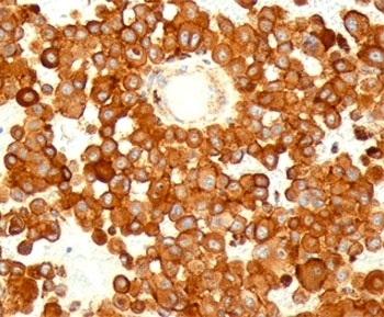
IHC testing of human melanoma stained with CD63 antibody (MX49.129.5).
IHC testing of mouse spleen stained with CD63 antibody (MX49.129.5).
FACS testing of human PBMC: Black = cells alone, Green = isotype control, Red = CD63 antibody PE conjugate.
FACS testing of mouse NIH3T3: Black = cells alone, Green = isotype control, Red = CD63 antibody PE conjugate.
Western blot testing of human spleen lysate with CD63 antibody at 2 ug/ml.
IHC testing of FFPE prostate carcinoma with CD63 antibody.
Immunofluorescence testing of HeLa cells with Alexa Fluor 488 conjugated CD63 antibody (green). F-actin filaments are labeled with Dylight 554 phalloidin (red), nuclei stained with DAPI (blue).
* Mehrwertsteuer und Versandkosten nicht enthalten. Irrtümer und Preisänderungen vorbehalten