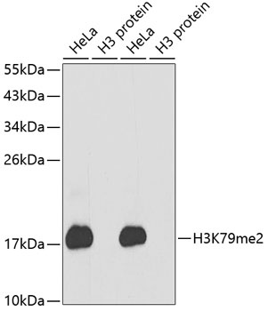
Western blot analysis of extracts of various cell lines, using DiMethyl-Histone H3-K79 antibody (orb1258180). Secondary antibody: HRP Goat Anti-Rabbit IgG (H+L) at 1:10000 dilution. Lysates/proteins: 25 ug per lane. Blocking buffer: 3% nonfat dry milk in TBST.
Dot-blot analysis of all sorts of methylation peptides using DiMethyl-Histone H3-K79 antibody (orb1258180).
Immunohistochemistry of paraffin-embedded human mammary cancer using DiMethyl-Histone H3-K79 antibody (orb1258180) at dilution of 1:200 (40x lens).
Immunohistochemistry of paraffin-embedded rat testis using DiMethyl-Histone H3-K79 antibody (orb1258180) at dilution of 1:200 (40x lens).
Immunohistochemistry of paraffin-embedded mouse testis using DiMethyl-Histone H3-K79 antibody (orb1258180) at dilution of 1:200 (40x lens).
Immunofluorescence analysis of 293T cells using DiMethyl-Histone H3-K79 antibody (orb1258180). Blue: DAPI for nuclear staining.
Chromatin Immunoprecipitation analysis of gamma-actin gene from 293 cell line, using DiMethyl-Histone H3-K79 antibody (orb1258180) and rabbit IgG. P1, P2 and P3 were probes located on gamma-actin gene as the schematic diagram illustrated. The amount of immunoprecipitated DNA was checked by quantitative PCR. Histogram was constructed by the ratios of the immunoprecipitated DNA to the input.
* Mehrwertsteuer und Versandkosten nicht enthalten. Irrtümer und Preisänderungen vorbehalten