
H2B K15ac Antibody, Unconjugated, Rabbit, Polyclonal
Artikelnummer:
BYT-ORB1259420
- Bilder (8)


| Artikelname: | H2B K15ac Antibody, Unconjugated, Rabbit, Polyclonal |
| Artikelnummer: | BYT-ORB1259420 |
| Hersteller Artikelnummer: | orb1259420 |
| Alternativnummer: | BYT-ORB1259420-100 |
| Hersteller: | Biorbyt |
| Wirt: | Rabbit |
| Kategorie: | Antikörper |
| Applikation: | IF, IHC, IP, WB |
| Spezies Reaktivität: | Human, Mouse |
| Immunogen: | AA synthetic acetylated peptide corresponding to residues surrounding K15 of human Histone H2B |
| Konjugation: | Unconjugated |
| H2B K15ac Antibody |
| Klonalität: | Polyclonal |
| Konzentration: | batch dependent |
| Puffer: | PBS with 0.02% sodium azide, 50% glycerol, pH 7.3. |
| Formulierung: | Liquid |
| Target-Kategorie: | H2B K15ac |
| Anwendungsbeschreibung: | Application Notes: WB: 1:500 - 1:2000IHC: 1:50 - 1:200IF: 1:50 - 1:100IP: 1:50 - 1:200 |
|
|
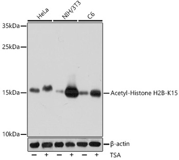
Western blot analysis of extracts of various cell lines, using Acetyl-Histone H2B-K15 antibody (orb1259420) at 1:1000 dilution. HeLa cells were treated by TSA (1 uM) at 37 °C for 18 hours. NIH/3T3 cells were treated by TSA (1 uM) at 37 °C for 18 hours. C6 cells were treated by TSA (1 uM) at 37 °C for 18 hours. Secondary antibody: HRP Goat Anti-Rabbit IgG (H+L) at 1:10000 dilution. Lysates/proteins: 25 ug per lane. Blocking buffer: 3% BSA. Detection: ECL Basic Kit. Exposure time: 10s. |
|
|
Immunohistochemistry of paraffin-embedded rat lung using H2B K15ac antibody (orb1259420) at dilution of 1:100 (40x lens). |
|
|
Immunohistochemistry of paraffin-embedded human breast cancer using H2B K15ac antibody (orb1259420) at dilution of 1:100 (40x lens). |
|
|
Immunohistochemistry of paraffin-embedded human gastric cancer using H2B K15ac antibody (orb1259420) at dilution of 1:100 (40x lens). |
|
|
Immunohistochemistry of paraffin-embedded mouse brain using H2B K15ac antibody (orb1259420) at dilution of 1:100 (40x lens). |
|
|
Immunofluorescence analysis of C6 cells using Acetyl-Histone H2B-K15 antibody (orb1259420) at dilution of 1:100. C6 cells were treated by TSA (1 uM) at 37 °C for 18 hours. Blue: DAPI for nuclear staining. |
|
|
Immunofluorescence analysis of HeLa cells using Acetyl-Histone H2B-K15 antibody (orb1259420) at dilution of 1:100. HeLa cells were treated by TSA (1 uM) at 37 °C for 18 hours. Blue: DAPI for nuclear staining. |
|
|
Immunofluorescence analysis of NIH/3T3 cells using Acetyl-Histone H2B-K15 antibody (orb1259420) at dilution of 1:100. NIH/3T3 cells were treated by TSA (1 uM) at 37 °C for 18 hours. Blue: DAPI for nuclear staining. |
Produktgarantie und fachkundiger Support
