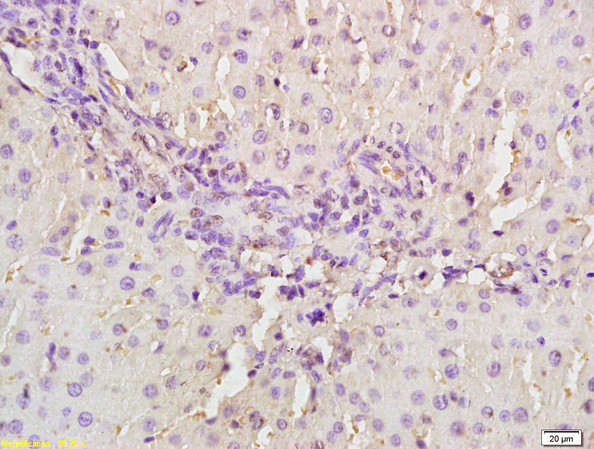
Immunohistochemical staining of rat liver using MASP2 antibody.
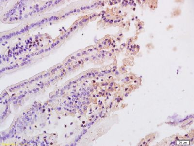
Immunohistochemical staining of rat ileum tissue using MASP2 antibody.
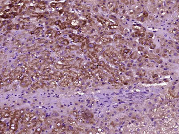
Paraformaldehyde-fixed, paraffin embedded (Human liver carcinoma), Antigen retrieval by boiling in sodium citrate buffer (pH6.0) for 15 min, Block endogenous peroxidase by 3% hydrogen peroxide for 20 minutes, Blocking buffer (normal goat serum) at 37C for 30 min, Antibody incubation with (MASP2) Polyclonal Antibody, Unconjugated (orb13562) at 1:400 overnight at 4C, followed by operating according to SP Kit (Rabbit) instructionsand DAB staining.
Paraformaldehyde-fixed, paraffin embedded (Human liver), Antigen retrieval by boiling in sodium citrate buffer (pH6.0) for 15 min, Block endogenous peroxidase by 3% hydrogen peroxide for 20 minutes, Blocking buffer (normal goat serum) at 37C for 30 min, Antibody incubation with (MASP2) Polyclonal Antibody, Unconjugated (orb13562) at 1:400 overnight at 4C, followed by operating according to SP Kit (Rabbit) instructionsand DAB staining.
Sample: Lung (Mouse) Lysate at 40 ug, Cerebrum (Mouse) Lysate at 40 ug Primary: Anti-MASP2 (orb13562) at 1/300 dilution, Secondary: IRDye800CW Goat Anti-Rabbit IgG at 1/20000 dilution, Predicted band size: 74 kD, Observed band size: 74 kD.
Sample: Plasma (Rat) Lysate at 40 ug, Primary: Anti-MASP2 (orb13562) at 1/1000 dilution, Secondary: IRDye800CW Goat Anti-Rabbit IgG at 1/20000 dilution, Predicted band size: 74 kD, Observed band size: 75 kD.
* Mehrwertsteuer und Versandkosten nicht enthalten. Irrtümer und Preisänderungen vorbehalten