E.coli-derived human PARK7 recombinant protein (Position: A2-D189). Human PARK7 shares 91% amino acid (aa) sequence identity with both mouse and rat PARK7.
Konjugation:
Unconjugated
Alternative Synonym:
DJ 1, DJ1, DJ-1, Oncogene DJ1, PARK7, PARK7, DJ-1, Parkinson disease protein 7, Protein DJ 1
Anti-PARK7/DJ1 Antibody (monoclonal, 4B10). Tested in Flow Cytometry, IF, IHC, ICC, WB applications. This antibody reacts with Human.
Klonalität:
Monoclonal
Konzentration:
Adding 0.2 ml of distilled water will yield a concentration of 500 µg/ml.
Each vial contains 4 mg Trehalose, 0.9 mg NaCl and 0.2 mg Na2HPO4.
Formulierung:
Lyophilized
Target-Kategorie:
Parkinson disease protein 7
Application Verdünnung:
Western blot, 0.25-0.5 µg/ml, Human Immunohistochemistry(Paraffin-embedded Section), 2-5 µg/ml, Human Immunocytochemistry/Immunofluorescence, 5 µg/ml, Human Flow Cytometry (Fixed), 1-3 µg/1x10 6 cells, Human
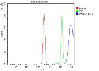
Flow Cytometry analysis of Hela cells using anti-PARK7/DJ1 antibody. Overlay histogram showing Hela cells (Blue line). To facilitate intracellular staining, cells were fixed with 4% paraformaldehyde and permeabilized with permeabilization buffer. The cells were blocked with 10% normal goat serum. And then incubated with mouse anti-PARK7/DJ1 Antibody (1 µg/1x10 6 cells) for 30 min at 20C. DyLight488 conjugated goat anti-mouse IgG (5-10 µg/1x10 6 cells) was used as secondary antibody for 30 minutes at 20C. Isotype control antibody (Green line) was mouse IgG (1 µg/1x10 6) used under the same conditions. Unlabelled sample without incubation with primary antibody and secondary antibody (Red line) was used as a blank control.
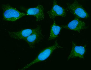
IF analysis of PARK7/DJ1 using anti-PARK7/DJ1 antibody. PARK7/DJ1 was detected in an immunocytochemical section of Hela cells. Enzyme antigen retrieval was performed using IHC enzyme antigen retrieval reagent for 15 mins. The cells were blocked with 10% goat serum. And then incubated with 5 µg/mL mouse anti-PARK7/DJ1 Antibody overnight at 4C. DyLight488 Conjugated Goat Anti-Mouse IgG was used as secondary antibody at 1:100 dilution and incubated for 30 minutes at 37C. The section was counterstained with DAPI. Visualize using a fluorescence microscope and filter sets appropriate for the label used.
IHC analysis of PARK7/DJ1 using anti-PARK7/DJ1 antibody. PARK7/DJ1 was detected in a paraffin-embedded section of human endometrial carcinoma tissue. Heat mediated antigen retrieval was performed in EDTA buffer (pH8.0, epitope retrieval solution). The tissue section was blocked with 10% goat serum. The tissue section was then incubated with 2 µg/ml mouse anti-PARK7/DJ1 Antibody overnight at 4C. Peroxidase Conjugated Goat Anti-mouse IgG was used as secondary antibody and incubated for 30 minutes at 37C. The tissue section was developed using HRP Conjugated Mouse IgG Super Vision Assay Kit with DAB as the chromogen.
IHC analysis of PARK7/DJ1 using anti-PARK7/DJ1 antibody. PARK7/DJ1 was detected in a paraffin-embedded section of human esophageal squamous carcinoma tissue. Heat mediated antigen retrieval was performed in EDTA buffer (pH8.0, epitope retrieval solution). The tissue section was blocked with 10% goat serum. The tissue section was then incubated with 2 µg/ml mouse anti-PARK7/DJ1 Antibody overnight at 4C. Peroxidase Conjugated Goat Anti-mouse IgG was used as secondary antibody and incubated for 30 minutes at 37C. The tissue section was developed using HRP Conjugated Mouse IgG Super Vision Assay Kit with DAB as the chromogen.
IHC analysis of PARK7/DJ1 using anti-PARK7/DJ1 antibody. PARK7/DJ1 was detected in a paraffin-embedded section of human liver cancer tissue. Heat mediated antigen retrieval was performed in EDTA buffer (pH8.0, epitope retrieval solution). The tissue section was blocked with 10% goat serum. The tissue section was then incubated with 2 µg/ml mouse anti-PARK7/DJ1 Antibody overnight at 4C. Peroxidase Conjugated Goat Anti-mouse IgG was used as secondary antibody and incubated for 30 minutes at 37C. The tissue section was developed using HRP Conjugated Mouse IgG Super Vision Assay Kit with DAB as the chromogen.
Western blot analysis of PARK7/DJ1 using anti-PARK7/DJ1 antibody. Electrophoresis was performed on a 5-20% SDS-PAGE gel at 70V (Stacking gel) / 90V (Resolving gel) for 2-3 hours. The sample well of each lane was loaded with 30 ug of sample under reducing conditions. Lane 1: human U20S whole cell lysates, Lane 2: human HepG2 whole cell lysates. After electrophoresis, proteins were transferred to a nitrocellulose membrane at 150 mA for 50-90 minutes. Blocked the membrane with 5% non-fat milk/TBS for 1.5 hour at RT. The membrane was incubated with mouse anti-PARK7/DJ1 antigen affinity purified monoclonal antibody at 0.5 µg/mL overnight at 4C, then washed with TBS-0.1% Tween 3 times with 5 minutes each and probed with a goat anti-mouse IgG-HRP secondary antibody at a dilution of
* Mehrwertsteuer und Versandkosten nicht enthalten. Irrtümer und Preisänderungen vorbehalten