Mouse monoclonal to GABA-A R Delta (HRP). The GABA-A receptor is a member of the superfamily of fast acting ligand-gated ion channels. The individual subunits of these receptors have similar sequences and structural features. GABA-A receptors are the major fast inhibitory neurotransmitter gated ion channels in the brain..
Application Notes: 2 µg/ml of SMC-345 was sufficient for detection of Delta1 GABA-A receptor in 10 µg of rat brain lysate by colorimetric immunoblot analysis using goat anti-mouse IgG:HRP as the secondary antibody
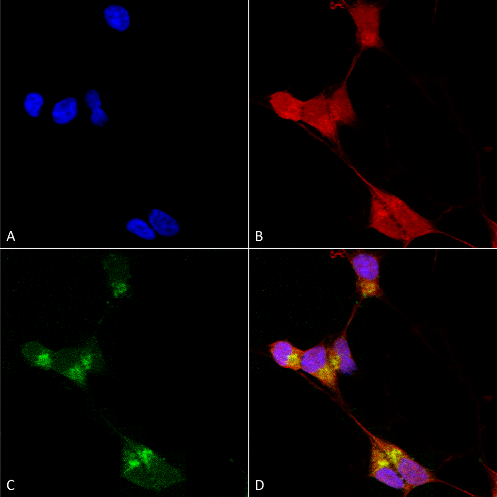
Immunocytochemistry/Immunofluorescence analysis using Mouse Anti-GABA-A Receptor Delta Monoclonal Antibody, Clone N151/3. Tissue: Neuroblastoma cells (SH-SY5Y). Species: Human. Fixation: 4% PFA for 15 min. Primary Antibody: Mouse Anti-GABA-A Receptor Delta Monoclonal Antibody at 1:100 for overnight at 4C with slow rocking. Secondary Antibody: AlexaFluor 488 at 1:1000 for 1 hour at RT. Counterstain: Phalloidin-iFluor 647 (red) F-Actin stain, Hoechst (blue) nuclear stain at 1:800, 1.6mM for 20 min at RT. (A) Hoechst (blue) nuclear stain. (B) Phalloidin-iFluor 647 (red) F-Actin stain. (C) GABA-A Receptor Delta Antibody (D) Composite.
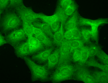
Immunocytochemistry/Immunofluorescence analysis using Mouse Anti-GABA A Receptor Monoclonal Antibody, Clone N151/3. Tissue: HaCaT cells. Species: Human. Fixation: Cold 100% methanol for 10 minutes at -20C. Primary Antibody: Mouse Anti-GABA A Receptor Monoclonal Antibody at 1:100 for 1 hour at RT. Secondary Antibody: FITC Goat Anti-Mouse (green) at 1:50 for 1 hour at RT. Localization: Diffuse cytoplasm and dull nuclei.
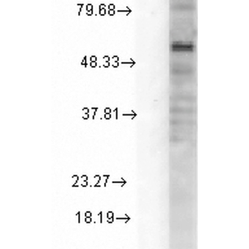
Western Blot analysis of Rat Cell line lysates showing detection of GABA A Receptor protein using Mouse Anti-GABA A Receptor Monoclonal Antibody, Clone N151/3. Load: 15 µg. Block: 1.5% BSA for 30 minutes at RT. Primary Antibody: Mouse Anti-GABA A Receptor Monoclonal Antibody at 1:1000 for 2 hours at RT. Secondary Antibody: Sheep Anti-Mouse IgG: HRP for 1 hour at RT.
Immunocytochemistry/Immunofluorescence analysis of Neuroblastom
Western Blot analysis of Rat Cell line lysates using GABA-A Receptor Delta antibody
* Mehrwertsteuer und Versandkosten nicht enthalten. Irrtümer und Preisänderungen vorbehalten