Anti PRTN3 antibody, anti proteinase 3 antibody, anti ACPA antibody, anti AGP7 antibody, anti C-ANCA antibody, anti CANCA antibody, anti MBN antibody, anti MBT antibody, anti NP4 antibody, anti P29 antibody, anti PR-3 antibody, anti PR3 antibody, anti C-ANCA antigen antibody, anti NP-4 antibody, anti Wegener granulomatosis autoantigen antibody, anti azurophil granule protein 7 antibody, anti leukocyte proteinase 3 antibody, anti myeloblastin antibody, anti neutrophil proteinase 4 antibody, anti serine proteinase antibody, anti neutrophil antibody, anti wegener autoantigen antibody
Goat polyclonal antibody to PRTN3.
Klonalität:
Polyclonal
Molekulargewicht:
27,8
Puffer:
Supplied at 0.5 mg/ml in Tris saline, 0.02% sodium azide, pH 7.3 with 0.5% bovine serum albumin. Aliquot and store at -20C. Minimize freezing and thawing.
Sequenz:
HNVRTQEPTQQ
Target-Kategorie:
proteinase 3 / myeloblastin (aa88-98)
Application Verdünnung:
ELISA: 1:8000, WB: 0.3-1 µg/ml, IHC-P: 5 µg/ml
Anwendungsbeschreibung:
Application Notes: WB: Approx 28kDa band observed in Human Spleen lysates (calculated MW of 27.8kDa according to NP_002768.3). Recommended concentration:0.3-1µg/ml. Primary incubation was 1 hour. IHC: Paraffin embedded Human Spleen. Recommended concentration: 5µg/ml
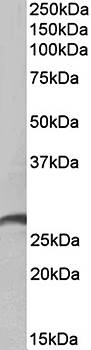
Primary incubation 1 hour at room temperature. Images A and B: Human Bone Marrow and Spleen lysate at primary Ab concentration 1 ug/ml. (Loaded 35 µg protein in RIPA buffer, per lane). Detected by chemiluminescence.
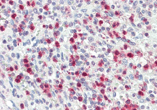
5 µg/ml staining of paraffin embedded Human Spleen. Steamed antigen retrieval with citrate buffer pH6, AP-staining.
Immunofluorescence analysis of paraformaldehyde fixed A431 cells, permeabilized with 0.15% Triton. Primary incubation 1hr (10 ug/ml) followed by Alexa Fluor 488 secondary antibody (2 ug/ml), showing cytoplasmic staining. The nuclear stain is DAPI (blue). Negative control: Unimmunized goat IgG (10 ug/ml) followed by Alexa Fluor 488 secondary antibody (2 ug/ml).
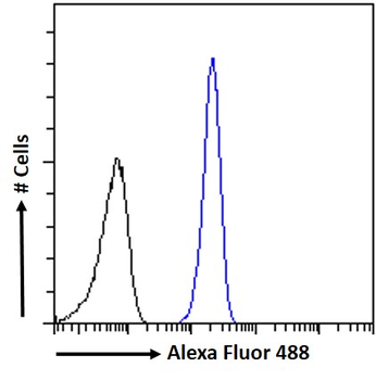
Flow cytometric analysis of paraformaldehyde fixed A431 cells (blue line), permeabilized with 0.5% Triton. Primary incubation 1hr (10 ug/ml) followed by Alexa Fluor 488 secondary antibody (1 ug/ml). IgG control: Unimmunized goat IgG (black line) followed by Alexa Fluor 488 secondary antibody.
Immunohistochemical staining of Human Spleen using PRTN3 antibody
Western blot analysis of Human Spleen lysate (35ug protein in RIPA buffer) using PRTN3 antibody
* Mehrwertsteuer und Versandkosten nicht enthalten. Irrtümer und Preisänderungen vorbehalten