E.coli-derived human Islet 1/ISL1 recombinant protein (Position: D118-E161). Human ISL1 shares 100% amino acid (aa) sequence identity with both mouse and rat ISL1.
Konjugation:
Unconjugated
Alternative Synonym:
Isl 1, ISL LIM homeobox 1, ISL1, Islet 1, ISLET1
Anti-Islet 1/ISL1 Antibody. Tested in ELISA, IF, IHC, ICC, WB applications. This antibody reacts with Human, Mouse, Rat.
Klonalität:
Polyclonal
Konzentration:
Adding 0.2 ml of distilled water will yield a concentration of 500 µg/ml.
Western blot, 0.25-0.5 µg/ml, Human Immunohistochemistry (Paraffin-embedded Section), 2-5 µg/ml, Mouse, Rat Immunocytochemistry/Immunofluorescence, 5 µg/ml, Human Immunofluorescence, 5 µg/ml, Mouse, Rat ELISA, 0.1-0.5 µg/ml, -
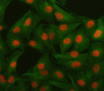
IF analysis of Islet 1/ISL1 using anti-Islet 1/ISL1 antibody and anti-Beta Tubulin antibody. Islet 1/ISL1 was detected in immunocytochemical section of A549 cell. Enzyme antigen retrieval was performed using IHC enzyme antigen retrieval reagent for 15 mins. The cells were blocked with 10% goat serum. And then incubated with 5 µg/mL rabbit anti-Islet 1/ISL1 Antibody and mouse anti-Beta Tubulin antibody overnight at 4C. Cy3 Conjugated Goat Anti-Rabbit IgG and DyLight488 Conjugated Goat Anti-Mouse IgG were used as secondary antibody at 1:500 dilution and incubated for 30 minutes at 37C. Visualize using a fluorescence microscope and filter sets appropriate for the label used.
IF analysis of Islet 1/ISL1 using anti-Islet 1/ISL1 antibody. Islet 1/ISL1 was detected in a paraffin-embedded section of mouse brain tissue. Heat mediated antigen retrieval was performed in EDTA buffer (pH8.0, epitope retrieval solution). The tissue section was blocked with 10% goat serum. The tissue section was then incubated with 5 µg/mL rabbit anti-Islet 1/ISL1 Antibody overnight at 4C. FITC Conjugated Goat Anti-Rabbit IgG was used as secondary antibody at 1:500 dilution and incubated for 30 minutes at 37C. Visualize using a fluorescence microscope and filter sets appropriate for the label used.
IF analysis of Islet 1/ISL1 using anti-Islet 1/ISL1 antibody. Islet 1/ISL1 was detected in a paraffin-embedded section of rat brain tissue. Heat mediated antigen retrieval was performed in EDTA buffer (pH8.0, epitope retrieval solution). The tissue section was blocked with 10% goat serum. The tissue section was then incubated with 5 µg/mL rabbit anti-Islet 1/ISL1 Antibody overnight at 4C. FITC Conjugated Goat Anti-Rabbit IgG was used as secondary antibody at 1:500 dilution and incubated for 30 minutes at 37C. Visualize using a fluorescence microscope and filter sets appropriate for the label used.
IHC analysis of Islet 1/ISL1 using anti-Islet 1/ISL1 antibody. Islet 1/ISL1 was detected in a paraffin-embedded section of mouse brain tissue. Heat mediated antigen retrieval was performed in EDTA buffer (pH8.0, epitope retrieval solution). The tissue section was blocked with 10% goat serum. The tissue section was then incubated with 2 µg/ml rabbit anti-Islet 1/ISL1 Antibody overnight at 4C. Peroxidase Conjugated Goat Anti-rabbit IgG was used as secondary antibody and incubated for 30 minutes at 37C. The tissue section was developed using HRP Conjugated Rabbit IgG Super Vision Assay Kit with DAB as the chromogen.
IHC analysis of Islet 1/ISL1 using anti-Islet 1/ISL1 antibody. Islet 1/ISL1 was detected in a paraffin-embedded section of rat brain tissue. Heat mediated antigen retrieval was performed in EDTA buffer (pH8.0, epitope retrieval solution). The tissue section was blocked with 10% goat serum. The tissue section was then incubated with 2 µg/ml rabbit anti-Islet 1/ISL1 Antibody overnight at 4C. Peroxidase Conjugated Goat Anti-rabbit IgG was used as secondary antibody and incubated for 30 minutes at 37C. The tissue section was developed using HRP Conjugated Rabbit IgG Super Vision Assay Kit with DAB as the chromogen.
Western blot analysis of Islet 1/ISL1 using anti-Islet 1/ISL1 antibody. Electrophoresis was performed on a 5-20% SDS-PAGE gel at 70V (Stacking gel) / 90V (Resolving gel) for 2-3 hours. The sample well of each lane was loaded with 30 ug of sample under reducing conditions. Lane 1: human SH-SY5Y whole cell lysates, Lane 2: human HepG2 whole cell lysates. After electrophoresis, proteins were transferred to a nitrocellulose membrane at 150 mA for 50-90 minutes. Blocked the membrane with 5% non-fat milk/TBS for 1.5 hour at RT. The membrane was incubated with rabbit anti-Islet 1/ISL1 antigen affinity purified polyclonal antibody at 0.5 µg/mL overnight at 4C, then washed with TBS-0.1% Tween 3 times with 5 minutes each and probed with a goat anti-rabbit IgG-HRP secondary antibody at a dilution of 1:5000 for 1.5 hour at RT. The signal is developed usi
* Mehrwertsteuer und Versandkosten nicht enthalten. Irrtümer und Preisänderungen vorbehalten