

GRP78 Antibody, Unconjugated, Rabbit, Polyclonal
Artikelnummer:
BYT-ORB1822447
- Bilder (5)


| Artikelname: | GRP78 Antibody, Unconjugated, Rabbit, Polyclonal |
| Artikelnummer: | BYT-ORB1822447 |
| Hersteller Artikelnummer: | orb1822447 |
| Alternativnummer: | BYT-ORB1822447-200 |
| Hersteller: | Biorbyt |
| Wirt: | Rabbit |
| Kategorie: | Antikörper |
| Applikation: | ICC, IF, IHC, IP, WB |
| Spezies Reaktivität: | Bovine, Canine, Frog, Fungi, Hamster, Human, Monkey, Mouse, Plant, Rabbit, Rat |
| Immunogen: | Rat GRP78 (Bip) synthetic peptide conjugated to KLH |
| Konjugation: | Unconjugated |
| Alternative Synonym: | GRP78, GRP-78, Grp78, GRP 78, HSPA5, HSPA 5, Heat shock 70 kDa protein 5, Heat Shock 70kDa Protein 5, BiP, BIP, Binding immunoglobulin protein, Immunoglobulin heavy chain-binding protein, Immunoglobulin Heavy Chain Binding Protein, Endoplasmic reticulum lumenal Ca(2+)-binding protein grp78, Endoplasmic reticulum lumenal, Ca2+ binding protein grp78, Glucose Regulated Protein 78kDa, 78 kDa glucose-regulated protein, 78 kDa glucose regulated protein, HSCe70, MIF2, mBiP, Sez7, AL022860, AU019543, D2Wsu141e, D2Wsu17e, FLJ26106, GRP78_HUMAN |
| Rabbit Anti-Rat GRP78 Polyclonal |
| Application Verdünnung: | WB (1:1000), ICC/IF (1:100), optimal dilutions for assays should be determined by the user. |
| Anwendungsbeschreibung: | Application Notes: A 1:1000 dilution was sufficient for detection of Grp78 in 10 µg of rat tissue lysate by ECL immunoblot analysis |
|
|
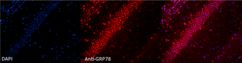
Immunocytochemistry/Immunofluorescence analysis using Rabbit Anti-GRP78 Polyclonal Antibody. Tissue: Hippocampal Section. Species: Mouse. Fixation: 4% Formaldehyde for 12 hours at RT. Paraffin embedded. Primary Antibody: Rabbit Anti-GRP78 Polyclonal Antibody at 1:100 for 12 hours at 4C. Secondary Antibody: Alexa Fluor 555 Goat Anti-Rabbit at 1:250 for 1 hour at RT. Counterstain: Hoechst at 1:1000 for 10 min at RT. Localization: Grp78 staining in mouse pyramidal cell layer. Magnification: 20x. Slice thickness: 7 microns. |
|
|
Western blot analysis of Human, Dog, Mouse Cell line lysates showing detection of GRP78 protein using Rabbit Anti-GRP78 Polyclonal Antibody. Primary Antibody: Rabbit Anti-GRP78 Polyclonal Antibody at 1:1000. |
|
|
Immunocytochemistry/Immunofluorescence analysis using Rabbit Anti-GRP78 Polyclonal Antibody. Tissue: Heat Shocked Cervical cancer cell line (HeLa). Species: Human. Fixation: 2% Formaldehyde for 20 min at RT. Primary Antibody: Rabbit Anti-GRP78 Polyclonal Antibody at 1:100 for 12 hours at 4C. Secondary Antibody: FITC Goat Anti-Rabbit (green) at 1:200 for 2 hours at RT. Counterstain: DAPI (blue) nuclear stain at 1:40000 for 2 hours at RT. Localization: Endoplasmic reticulum lumen. Melanosome. Cytoplasm. Magnification: 100x. (A) DAPI (blue) nuclear stain. (B) Anti-GRP78 Antibody. (C) Composite. Heat Shocked at 42C for 30 min. |
|
|
Western blot analysis of Rat Tissue lysates showing detection of GRP78 protein using Rabbit Anti-GRP78 Polyclonal Antibody. Load: 15 µgprotein. Block: 1.5% BSA. Primary Antibody: Rabbit Anti-GRP78 Polyclonal Antibody at 1:1000 for 2 hours at RT. Secondary Antibody: Donkey Anti-Rabbit IgG: HRP for 1 hour at RT. |
|
|
Immunocytochemistry/Immunofluorescence analysis using Rabbit Anti-GRP78 Polyclonal Antibody. Tissue: Heat Shocked Cervical cancer cell line (HeLa). Species: Human. Fixation: 2% Formaldehyde for 20 min at RT. Primary Antibody: Rabbit Anti-GRP78 Polyclonal Antibody at 1:100 for 12 hours at 4C. Secondary Antibody: APC Goat Anti-Rabbit (red) at 1:200 for 2 hours at RT. Counterstain: DAPI (blue) nuclear stain at 1:40000 for 2 hours at RT. Localization: Endoplasmic reticulum lumen. Melanosome. Cytoplasm. Magnification: 20x. (A) DAPI (blue) nuclear stain. (B) Anti-GRP78 Antibody. (C) Composite. Heat Shocked at 42C for 30 min. |
Produktgarantie und fachkundiger Support
