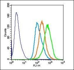
Flow cytometric analysis of Hela cells using COX4 antibody.
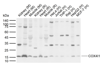
Western blot analysis of extracts from kidney using COX4 antibody.
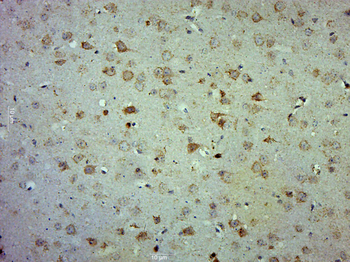
Paraformaldehyde-fixed, paraffin embedded (Mouse brain), Antigen retrieval by boiling in sodium citrate buffer (pH6.0) for 15 min, Block endogenous peroxidase by 3% hydrogen peroxide for 20 minutes, Blocking buffer (normal goat serum) at 37C for 30 min, Antibody incubation with (COX4) Polyclonal Antibody, Unconjugated (orb182884) at 1:500 overnight at 4C, followed by a conjugated secondary for 20 minutes and DAB staining.
Sample: Lane 1: Mouse Kidney tissue lysates, Lane 2: Mouse Heart tissue lysates, Lane 3: Mouse Muscle tissue lysates, Lane 4: Rat Kidney tissue lysates, Lane 5: Rat Heart tissue lysates, Lane 6: Rat Muscle tissue lysates, Lane 7: Human HeLa cell lysates, Lane 8: Human Jurkat cell lysates, Lane 9: Human THP-1 cell lysates, Lane 10: Human Raji cell lysates, Lane 11: Human HepG2 cell lysates, Lane 12: Human MCF-7 cell lysates, Primary: Anti-COX4I1 (orb182884) at 1/1000 dilution, Secondary: IRDye800CW Goat Anti-Rabbit IgG at 1/20000 dilution, Predicted band size: 17 kDa, Observed band size: 16 kDa.
Tissue/Cell: Rat brain tissue, 4% Paraformaldehyde-fixed and paraffin-embedded, Antigen retrieval: citrate buffer (0.01M, pH 6.0), Boiling bathing for 15 min, Block endogenous peroxidase by 3% Hydrogen peroxide for 30 min, Blocking buffer (normal goat serum) at 37C for 20 min, Incubation: Anti-COX4 Polyclonal Antibody, Unconjugated (orb182884) 1:200, overnight at 4C, followed by conjugation to the secondary antibody and DAB staining.
* Mehrwertsteuer und Versandkosten nicht enthalten. Irrtümer und Preisänderungen vorbehalten