Purified polyclonal antibody supplied in PBS with 0.09% (W/V) sodium azide. This antibody is purified through a protein A column, followed by peptide affinity purification.
Application Verdünnung:
IHC-P-Leica - 1:500, IF - 1:25, WB - 1:2000
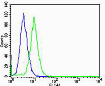
Flow cytometric analysis of Hela cells using GLI2 Antibody (C-term) (green) compared to an isotype control of rabbit IgG (blue). Diluted at 1:25 dilution. An Alexa Fluor 488 goat anti-rabbit lgG at 1:400 dilution was used as the secondary antibody.
Western blot analysis of lysates from 293, Jurkat cell line (from left to right), using GLI2 Antibody (C-term). Diluted at 1:1000 at each lane. A goat anti-rabbit IgG H&L (HRP) at 1:5000 dilution was used as the secondary antibody. Lysates at 35 ug per lane.
All lanes: Anti-GLI2 Antibody (C-term) at 1:2000 dilution. Lane 1: Jurkat whole cell lysate. Lane 2: CCRF-CEM whole cell lysate. Lysates/proteins at 20 µg per lane. Secondary Goat Anti-Rabbit IgG, (H+L), Peroxidase conjugated at 1/10000 dilution. Predicted band size: 168 kDa. Blocking/Dilution buffer: 5% NFDM/TBST.
Immunohistochemical analysis of paraffin-embedded Human kidney tissue was performed on the Leica BOND RXm. Tissue was fixed with formaldehyde at room temperature, antigen retrieval was by heat mediation with a EDTA buffer (pH9.0). Samples were incubated with primary antibody (1:500) for 1 hours at room temperature. A undiluted biotinylated CRF Anti-Polyvalent HRP Polymer antibody was used as the secondary antibody.
Immunohistochemical analysis of paraffin-embedded Human breast carcinoma tissue was performed on the Leica BOND RXm. Tissue was fixed with formaldehyde at room temperature, antigen retrieval was by heat mediation with a EDTA buffer (pH9.0). Samples were incubated with primary antibody (1:500) for 1 hours at room temperature. A undiluted biotinylated CRF Anti-Polyvalent HRP Polymer antibody was used as the secondary antibody.
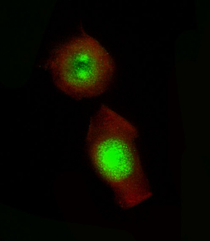
Immunofluorescent analysis of 4% paraformaldehyde-fixed, 0.1% Triton X-100 permeabilized PC-3 cells labeling GLI2 at 1/25 dilution, followed by Dylight 488-conjugated goat anti-Rabbit IgG secondary antibody at 1/200 dilution (green). Immunofluorescence image showing Nucleus and Weak Cytoplasm staining on PC-3 cell line. Cytoplasmic actin is detected with Dylight 554 Phalloidin (red). The nuclear counter stain is DAPI (blue).
* Mehrwertsteuer und Versandkosten nicht enthalten. Irrtümer und Preisänderungen vorbehalten