NKG2-A/NKG2-B type II integral membrane protein, CD159 antigen-like family member A, NK cell receptor A, NKG2-A/B-activating NK receptor, CD159a, KLRC1, NKG2A
Purified polyclonal antibody supplied in PBS with 0.09% (W/V) sodium azide. This antibody is purified through a protein A column, followed by peptide affinity purification.
Target-Kategorie:
This KLRC1 antibody is generated from rabbits immunized with a KLH conjugated synthetic peptide between 180-206 amino acids from the C-terminal region of human KLRC1.
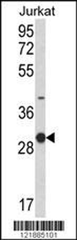
Western blot analysis of KLRC1 Antibody (C-term) in Jurkat cell line lysates (35 ug/lane). KLRC1 (arrow) was detected using the purified Pab.
Anti-KLRC1 Antibody (C-term) at 1:2000 dilution + Jurkat whole cell lysate. Lysates/proteins at 20 µg per lane. Secondary Goat Anti-Rabbit IgG, (H+L), Peroxidase conjugated at 1/10000 dilution. Predicted band size: 26 kDa. Blocking/Dilution buffer: 5% NFDM/TBST.
Formalin-fixed and paraffin-embedded human kidney carcinoma with KLRC1 Antibody (C-term), which was peroxidase-conjugated to the secondary antibody, followed by DAB staining. This data demonstrates the use of this antibody for immunohistochemistry, clinical relevance has not been evaluated.
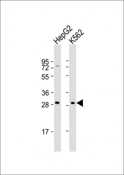
All lanes: Anti-KLRC1 Antibody (C-term) at 1:2000 dilution. Lane 1: HepG2 whole cell lysates. Lane 2: K562 whole cell lysates. Lysates/proteins at 20 µg per lane. Secondary Goat Anti-Rabbit IgG, (H+L), Peroxidase conjugated at 1/10000 dilution. Predicted band size: 26 kDa. Blocking/Dilution buffer: 5% NFDM/TBST.
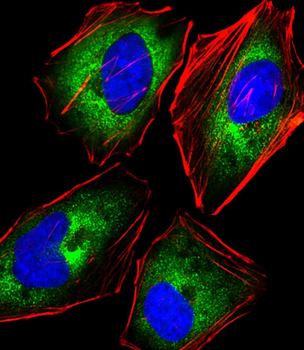
Immunofluorescent analysis of 4% paraformaldehyde-fixed, 0.1% Triton X-100 permeabilized HeLa (human cervical epithelial adenocarcinoma cell line) cells labeling KLRC1 at 1/25 dilution, followed by Dylight 488-conjugated goat anti-rabbit IgG secondary antibody at 1/200 dilution (green). Immunofluorescence image showing cytoplasm staining on HeLa cell line. Cytoplasmic actin is detected with Dylight 554 Phalloidin at 1/100 dilution (red).The nuclear counter stain is DAPI (blue).
Overlay histogram showing Jurkat cells stained (green line). The cells were fixed with 2% paraformaldehyde (10 min). The cells were then icubated in 2% bovine serum albumin to block non-specific protein-protein interactions followed by the antibody (1:25 dilution) for 60 min at 37C. The secondary antibody used was Goat-Anti-Rabbit IgG, DyLight 488 Conjugated Highly Cross-Adsorbed at 1/400 dilution for 40 min at 37C. Isotype control antibody (blue line) was rabbit IgG (1 µg/1x10 6 cells) used under the same conditions. Acquisition of > 10000 events was performed.
* Mehrwertsteuer und Versandkosten nicht enthalten. Irrtümer und Preisänderungen vorbehalten