Purified polyclonal antibody supplied in PBS with 0.09% (W/V) sodium azide. This antibody is purified through a protein A column, followed by peptide affinity purification.
Application Verdünnung:
WB - 1:500
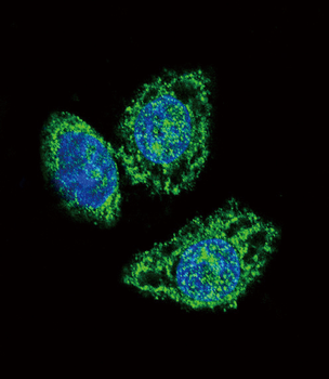
Confocal immunofluorescent analysis of DUSP6 Antibody (Center) with Hela cell followed by Alexa Fluor 488-conjugated goat anti-rabbit lgG (green). DAPI was used to stain the cell nuclear (blue).
All lanes: Anti-DUSP6 Antibody (Center) at 1:2000 dilution. Lane 1: Human brain lysate. Lane 2: Human lung lysate. Lane 3: Rat brain lysate. Lysates/proteins at 20 µg per lane. Secondary Goat Anti-Rabbit IgG, (H+L), Peroxidase conjugated at 1/10000 dilution. Predicted band size: 42 kDa. Blocking/Dilution buffer: 5% NFDM/TBST.
All lanes: Anti-DUSP6 Antibody (Center) at 1:2000 dilution. Lane 1: HepG2 whole cell lysate. Lane 2: Human brain tissue lysate. Lane 3: Rat brain tissue lysate. Lysates/proteins at 20 µg per lane. Secondary Goat Anti-Rabbit IgG, (H+L), Peroxidase conjugated at 1/10000 dilution. Predicted band size: 42 kDa. Blocking/Dilution buffer: 5% NFDM/TBST.
All lanes: Anti-DUSP6 Antibody (Center) at 1:500 dilution. Lane 1: Human brain tissue lysate. Lane 2: Mouse brain tissue lysate. Lane 3: Rat brain tissue lysate. Lysates/proteins at 20 µg per lane. Secondary Goat Anti-Rabbit IgG, (H+L), Peroxidase conjugated at 1/10000 dilution. Predicted band size: 42 kDa. Blocking/Dilution buffer: 5% NFDM/TBST.
Immunohistochemical analysis of paraffin-embedded human kidney tissue was performed on the Leica BOND RXm. Tissue was fixed with formaldehyde at room temperature, antigen retrieval was by heat mediation with a EDTA buffer (pH9.0). Samples were incubated with primary antibody (1:500) for 1 hours at room temperature. A undiluted biotinylated CRF Anti-Polyvalent HRP Polymer antibody was used as the secondary antibody.
Immunohistochemical analysis of paraffin-embedded human colon carcinoma tissue was performed on the Leica BOND RXm. Tissue was fixed with formaldehyde at room temperature, antigen retrieval was by heat mediation with a EDTA buffer (pH9.0). Samples were incubated with primary antibody (1:500) for 1 hours at room temperature. A undiluted biotinylated CRF Anti-Polyvalent HRP Polymer antibody was used as the secondary antibody.
* Mehrwertsteuer und Versandkosten nicht enthalten. Irrtümer und Preisänderungen vorbehalten