Purified polyclonal antibody supplied in PBS with 0.09% (W/V) sodium azide. This antibody is prepared by Saturated Ammonium Sulfate (SAS) precipitation followed by dialysis against PBS.
Application Verdünnung:
WB - 1:1000, IHC-P - 1:100-500, FC - 1:10-50, IF - 1:10-50
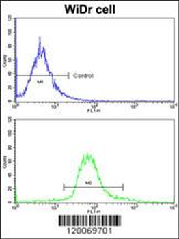
Flow cytometric analysis of widr cells using GLO1 Antibody (N-term) (bottom histogram) compared to a negative control cell (top histogram) FITC-conjugated goat-anti-rabbit secondary antibodies were used for the analysis.
Confocal immunofluorescent analysis of GLO1 Antibody (N-term) with WiDr cell followed by Alexa Fluor 488-conjugated goat anti-rabbit lgG (green). DAPI was used to stain the cell nuclear (blue).
Formalin-fixed and paraffin-embedded human brain tissue reacted with GLO1 Antibody (N-term), which was peroxidase-conjugated to the secondary antibody, followed by DAB staining. This data demonstrates the use of this antibody for immunohistochemistry, clinical relevance has not been evaluated.
Western blot analysis of GLO1 antibody (N-term) in HL60 cell line lysates (35 ug/lane). GLO1 (arrow) was detected using the purified Pab.
Western blot analysis of GLO1 (arrow) using rabbit polyclonal GLO1 Antibody (N-term). 293 cell lysates (2 ug/lane) either nontransfected (Lane 1) or transiently transfected (Lane 2) with the GLO1 gene.
All lanes: Anti-GLO1 Antibody (N-term) at 1:1000 dilution. Lane 1: Hela whole cell lysate. Lane 2: HepG2 whole cell lysate. Lysates/proteins at 20 µg per lane. Secondary Goat Anti-Rabbit IgG, (H+L), Peroxidase conjugated at 1/10000 dilution. Predicted band size: 21 kDa. Blocking/Dilution buffer: 5% NFDM/TBST.
* Mehrwertsteuer und Versandkosten nicht enthalten. Irrtümer und Preisänderungen vorbehalten