Purified polyclonal antibody supplied in PBS with 0.09% (W/V) sodium azide. This antibody is prepared by Saturated Ammonium Sulfate (SAS) precipitation followed by dialysis against PBS.
Application Verdünnung:
WB - 1:1000, IHC-P - 1:100-500, FC - 1:10-50
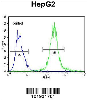
SEPT9 Antibody (A555) flow cytometric analysis of HepG2 cells (right histogram) compared to a negative control cell (left histogram). FITC-conjugated goat-anti-rabbit secondary antibodies were used for the analysis.
SEPT9 Antibody (A555) immunohistochemistry analysis in formalin fixed and paraffin embedded kidney tissue followed by peroxidase conjugation of the secondary antibody and DAB staining. This data demonstrates the use of SEPT9 Antibody (A555) for immunohistochemistry. Clinical relevance has not been evaluated.
Formalin-fixed and paraffin-embedded human cancer tissue reacted with the primary antibody, which was peroxidase-conjugated to the secondary antibody, followed by AEC staining. This data demonstrates the use of this antibody for immunohistochemistry, clinical relevance has not been evaluated. BC = breast carcinoma, HC = hepatocarcinoma.
The anti-SEPT9 Pab is used in Western blot to detect SEPT9 in Jurkat cell lysate.
Western blot analysis of lysates from A431, Hela cell line (from left to right), using SEPT9 Antibody (C-term). Diluted at 1:1000 at each lane. A goat anti-rabbit IgG H&L (HRP) at 1:5000 dilution was used as the secondary antibody. Lysates at 35 ug per lane.
Western blot analysis of lysates from human kidney and liver tissue lysate (from left to right), using SEPT9 Antibody (C-term). Diluted at 1:1000 at each lane. A goat anti-rabbit IgG H&L (HRP) at 1:5000 dilution was used as the secondary antibody. Lysates at 35 ug per lane.
* Mehrwertsteuer und Versandkosten nicht enthalten. Irrtümer und Preisänderungen vorbehalten