E.coli-derived human PARK7 recombinant protein (Position: A2-D189). Human PARK7 shares 91% amino acid (aa) sequence identity with both mouse and rat PARK7.
Konjugation:
Unconjugated
Alternative Synonym:
Protein deglycase DJ-1, DJ-1, 3.1.2.-, 3.5.1.-, Oncogene DJ1, Parkinson disease protein 7, PARK7
PARK7/DJ1 Rabbit Polyclonal Antibody
Klonalität:
Polyclonal
Konzentration:
Adding 0.2 ml of distilled water will yield a concentration of 500 µg/ml.
Each vial contains antibody formulated with stabilizing components, 0.9mg NaCl, 0.2mg Na2HPO4, 0.01mg NaN3. *This antibody is supplied in a stabilized formulation. Compatibility with conjugation reactions depends on the chemistry of the conjugation method
Formulierung:
Lyophilized
Target-Kategorie:
Parkinson disease protein 7
Application Verdünnung:
Western blot, 0.1-0.5µg/ml, Human, Mouse, Rat Immunohistochemistry (Paraffin-embedded Section), 0.5-1µg/ml, Human, Mouse, Rat Immunocytochemistry/Immunofluorescence, 5 µg/ml, Human Immunoprecipitation, 0.5-2 µg/ml, Human
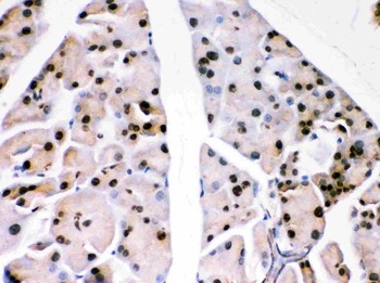
IHC(P) analysis of Mouse Pancreas Tissue using Anti-PARK7 Picoband antibody.
Anti-PARK7 Picoband antibody, IHC(P): Human Pancreatic Cancer Tissue.
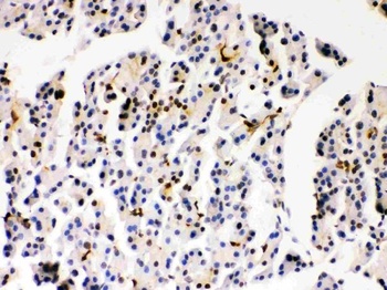
IHC(P) analysis of Rat Pancreas Tissue using Anti-PARK7 Picoband antibody.
Anti-PARK7 Picoband antibody, IHC(P): Rat Pancreas Tissue.
IHC analysis of PARK7 using anti-PARK7 antibody. PARK7 was detected in immunocytochemical section of A549 Cell. Enzyme antigen retrieval was performed using IHC enzyme antigen retrieval reagent for 15 mins. The cells were blocked with 10% goat serum. And then incubated with 1 µg/ml rabbit anti-PARK7 Antibody overnight at 4C. Biotinylated goat anti-rabbit IgG was used as secondary antibody and incubated for 30 minutes at 37C. The section was developed using Strepavidin-Biotin-Complex (SABC) with DAB as the chromogen.
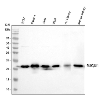
Western blot analysis of PARK7 using anti-PARK7 antibody. Electrophoresis was performed on a 5-20% SDS-PAGE gel at 70V (Stacking gel) / 90V (Resolving gel) for 2-3 hours. The sample well of each lane was loaded with 30 ug of sample under reducing conditions. Lane 1: human 293T whole cell lysates, Lane 2: human PANC-1 whole cell lysates, Lane 3: human Hela whole cell lysates, Lane 4: human U251 whole cell lysates, Lane 5: rat kidney tissue lysates, Lane 6: mouse kidney tissue lysates. After electrophoresis, proteins were transferred to a nitrocellulose membrane at 150 mA for 50-90 minutes. Blocked the membrane with 5% non-fat milk/TBS for 1.5 hour at RT. The membrane was incubated with rabbit anti-PARK7 antigen affinity purified polyclonal antibody at 0.5 µg/mL overnight at 4C, then washed with TBS-0.1% Tween 3 times with 5 minutes each and probed with a goat anti-rabbit IgG-HRP secondary antibody at a dilution of 1:5000 for 1.5 hour at RT. The signal is developed using an Enhanced Chemiluminescent detection (ECL) kit with Tanon 5200 system. A specific band was detected for PARK7 at approximately 20 kDa. The expected band size for PARK7 is at 20 kDa.
* Mehrwertsteuer und Versandkosten nicht enthalten. Irrtümer und Preisänderungen vorbehalten