A synthetic peptide corresponding to aa 202-216 (GTYQDVGSLNIADVQ) of human CD79a protein (JCB117 and HM47/A9) was used as the immunogen for this antibody cocktail.
Konjugation:
Unconjugated
A disulphide-linked heterodimer, consisting of mb-1 (or CD79a) and B29 (or CD79b) polypeptides, is non-covalently associated with membrane-bound immunoglobulins on B cells. This complex of mb-1 and B29 polypeptides and immunoglobulin constitute the B cell Ag receptor. CD79a first appears at pre B cell stage, early in maturation, and persists until the plasma cell stage where it is found as an intracellular component. CD79a is found in the majority of acute leukemias of precursor B cell type, in B cell lines, B cell lymphomas, and in some myelomas. It is not present in myeloid or T cell lines. CD79a antibody is generally used to complement CD20 antibody especially for mature B-cell lymphomas after treatment with Rituximab (anti-CD20). This antibody cocktail will stain many of the same lymphomas as CD20 antibody, but also is more likely to stain B-lymphoblastic lymphoma/leukemia than is CD20 antibody. CD79a antibody also stains more cases of plasma cell myeloma and occasionally some types of endothelial cells as well.
Klonalität:
Monoclonal
Klon-Bezeichnung:
[JCB117 + HM47/A9]
Puffer:
0.2 mg/ml in 1X PBS with 0.1 mg/ml rAlbumin and 0.05% sodium azide
Application Verdünnung:
Western blot: 1-2ug/ml,Flow cytometry: 1-2ug/million cells,Immunofluorescence: 1-2ug/ml,Immunohistochemistry (FFPE): 1-2ug/ml for 30 min at RT
Anwendungsbeschreibung:
Application Notes: The concentration stated for each application is a general starting point. Variations in protocols, secondaries and substrates may require the antibody to be titered up or down for optimal performance.1. Staining of formalin-fixed tissues requires boiling tissue sections in pH 9 10mM Tris with 1mM EDTA for 10-20 min followed by cooling at RT for 20 minutes.2. The prediluted format is supplied in a dropper bottle and is optimized for use in IHC. After epitope retrieval step (if required), drip mAb solution onto the tissue section and incubate at RT for 30 min
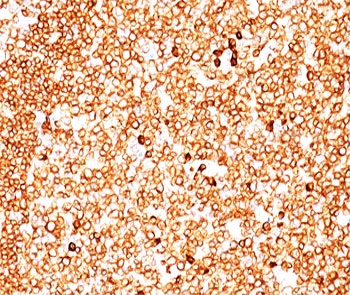
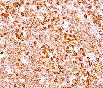
IHC testing of FFPE human tonsil (10X) stained with CD79a antibody cocktail (clone JCB117 + HM47/A9).
IHC testing of FFPE human tonsil (10X) stained with CD79a antibody cocktail (clone JCB117 + HM47/A9).
Flow cytometry testing of human Raji cells with CD79a antibody cocktail (clone JCB117 + HM47/A9), Red = isotype control, Blue = CD79a antibody cocktail.
Western blot testing of human Raji cell lysate with CD79a antibody cocktail (clone JCB117 + HM47/A9). Expected molecular weight: 25-47 kDa depending on glycosylation level.
Immunofluorescent staining of PFA-fixed human Raji cells with CD79a antibody cocktail (clone JCB117 + HM47/A9, green) and Reddot nuclear stain (red).
SDS-PAGE analysis of purified, BSA-free CD79a antibody cocktail (clone JCB117 + HM47/A9) as confirmation of integrity and purity.
* Mehrwertsteuer und Versandkosten nicht enthalten. Irrtümer und Preisänderungen vorbehalten