A cytoskeleton preparation containing Cytokeratin 8 was used as the immunogen for this antibody.
Konjugation:
Unconjugated
Cytokeratin 8 (CK8) belongs to the type II (or B or basic) subfamily of high molecular weight cytokeratins and exists in combination with cytokeratin 18 (CK18). Cytokeratin 8 is primarily found in the non-squamous epithelia and is present in majority of adenocarcinomas and ductal carcinomas. It is absent in squamous cell carcinomas. Hepatocellular carcinomas are defined by the use of antibody that recognizes only cytokeratin 8 and 18. Cytokeratin 8 exists on several types of normal and neoplastic epithelia, including many ductal and glandular epithelia such as colon, stomach, small intestine, trachea, and esophagus as well as in transitional epithelium. antibody to Cytokeratin 8 does not react with skeletal muscle or nerve cells. Epithelioid sarcoma, chordoma, and adamantinoma show strong positivity corresponding to that of simple epithelia (with antibodies against Cytokeratin 8, 18 and 19). Reportedly, Cytokeratin 8 antibody is useful for the differentiation of lobular (iring-like, perinucleari) from ductal (iperipheral-predominanti) carcinoma of the breast.
Klonalität:
Monoclonal
Puffer:
0.2 mg/ml in 1X PBS with 0.1 mg/ml rAlbumin and 0.05% sodium azide
Application Verdünnung:
Western blot: 1-2ug/ml,Flow cytometry: 1-2ug/million cells,Immunofluorescence: 1-2ug/ml,Immunohistochemistry (FFPE): 1-2ug/ml for 30 min at RT
Anwendungsbeschreibung:
Application Notes: The concentration stated for each application is a general starting point. Variations in protocols, secondaries and substrates may require the antibody to be titered up or down for optimal performance.1. Staining of formalin-fixed tissues requires boiling tissue sections in 10mM Citrate Buffer, pH 6.0, for 10-20 min followed by cooling at RT for 20 minutes.2. The prediluted format is supplied in a dropper bottle and is optimized for use in IHC. After epitope retrieval step (if required), drip mAb solution onto the tissue section and incubate at RT for 30 min
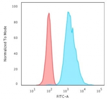
Flow cytometry testing of permeabilized human HeLa cells with Cytokeratin 8 antibody (clone H1), Red = isotype control, Blue = Cytokeratin 8 antibody.
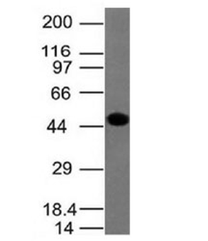
Western blot testing of human HCT-116 lysate with Cytokeratin 8 antibody.
Western blot testing of A431 lysate with Cytokeratin 8 antibody
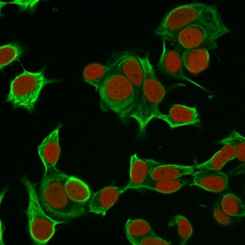
Immunofluorescent staining of permeabilized human MCF7 cells with Cytokeratin 8 antibody (clone H1, green) and Reddot nuclear stain (red).
Immunofluorescent staining of permeabilized human HCT-116 cells with Cytokeratin 8 antibody (clone H1, green) and Reddot nuclear stain (red).
IHC testing of human colon carcinoma stained with Cytokeratin 8 antibody
* Mehrwertsteuer und Versandkosten nicht enthalten. Irrtümer und Preisänderungen vorbehalten