Purified polyclonal antibody supplied in PBS with 0.09% (W/V) sodium azide. This antibody is prepared by Saturated Ammonium Sulfate (SAS) precipitation followed by dialysis against PBS.
Confocal immunofluorescent analysis of DARS Antibody (N-term) with HepG2 cell followed by Alexa Fluor 488-conjugated goat anti-rabbit lgG (green). DAPI was used to stain the cell nuclear (blue).
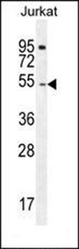
DARS Antibody (N-term) western blot analysis in Jurkat cell line lysates (35 ug/lane). This demonstrates the DARS antibody detected the DARS protein (arrow).
Western blot analysis of DARS (arrow) using rabbit polyclonal DARS Antibody (N-term). 293 cell lysates (2 ug/lane) either nontransfected (Lane 1) or transiently transfected (Lane 2) with the DARS gene.
DARS Antibody (N-term) immunohistochemistry analysis in formalin fixed and paraffin embedded human liver tissue followed by peroxidase conjugation of the secondary antibody and DAB staining. This data demonstrates the use of DARS Antibody (N-term) for immunohistochemistry. Clinical relevance has not been evaluated.
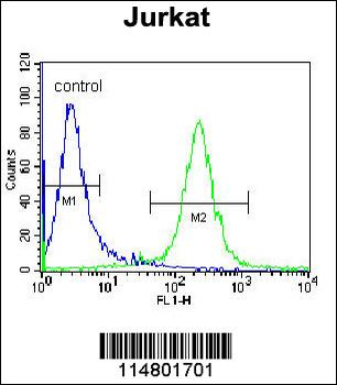
DARS Antibody (N-term) flow cytometric analysis of Jurkat cells (right histogram) compared to a negative control cell (left histogram). FITC-conjugated donkey-anti-rabbit secondary antibodies were used for the analysis.
orb29039
* Mehrwertsteuer und Versandkosten nicht enthalten. Irrtümer und Preisänderungen vorbehalten