A synthetic peptide corresponding to a sequence at the C-terminus of human Aquaporin 4, different from the related mouse sequence by two amino acids, and from the related rat sequence by three amino acids.
Konjugation:
Unconjugated
Alternative Synonym:
Aquaporin-4, AQP-4, Mercurial-insensitive water channel, MIWC, WCH4, AQP4
Aquaporin 4/AQP4 Rabbit Polyclonal Antibody
Klonalität:
Polyclonal
Konzentration:
Adding 0.2 ml of distilled water will yield a concentration of 500 µg/ml.
Each vial contains 4 mg Trehalose, 0.9 mg NaCl and 0.2 mg Na2HPO4.
Formulierung:
Lyophilized
Target-Kategorie:
Aquaporin-4
Application Verdünnung:
Western blot, 0.1-0.5µg/ml, Mouse, Rat Immunohistochemistry (Paraffin-embedded Section), 2-5µg/ml, Human, Mouse, Rat Immunohistochemistry (Frozen Section), 0.5-1µg/ml, Mouse, Rat
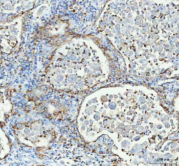
IHC analysis of Aquaporin 4 using anti-Aquaporin 4 antibody. Aquaporin 4 was detected in a paraffin-embedded section of human lung cancer tissue. Heat mediated antigen retrieval was performed in EDTA buffer (pH8.0, epitope retrieval solution). The tissue section was blocked with 10% goat serum. The tissue section was then incubated with 2 µg/ml rabbit anti-Aquaporin 4 Antibody overnight at 4C. Peroxidase Conjugated Goat Anti-rabbit IgG was used as secondary antibody and incubated for 30 minutes at 37C. The tissue section was developed using HRP Conjugated Rabbit IgG Super Vision Assay Kit with DAB as the chromogen.
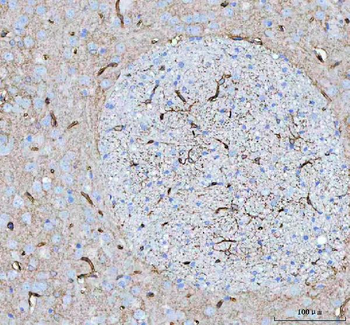
IHC analysis of Aquaporin 4 using anti-Aquaporin 4 antibody. Aquaporin 4 was detected in a paraffin-embedded section of mouse brain tissue. Heat mediated antigen retrieval was performed in EDTA buffer (pH8.0, epitope retrieval solution). The tissue section was blocked with 10% goat serum. The tissue section was then incubated with 2 µg/ml rabbit anti-Aquaporin 4 Antibody overnight at 4C. Peroxidase Conjugated Goat Anti-rabbit IgG was used as secondary antibody and incubated for 30 minutes at 37C. The tissue section was developed using HRP Conjugated Rabbit IgG Super Vision Assay Kit with DAB as the chromogen.
IHC analysis of Aquaporin 4 using anti-Aquaporin 4 antibody. Aquaporin 4 was detected in a paraffin-embedded section of rat brain tissue. Heat mediated antigen retrieval was performed in EDTA buffer (pH8.0, epitope retrieval solution). The tissue section was blocked with 10% goat serum. The tissue section was then incubated with 2 µg/ml rabbit anti-Aquaporin 4 Antibody overnight at 4C. Peroxidase Conjugated Goat Anti-rabbit IgG was used as secondary antibody and incubated for 30 minutes at 37C. The tissue section was developed using HRP Conjugated Rabbit IgG Super Vision Assay Kit with DAB as the chromogen.
IHC analysis of Aquaporin 4 using anti-Aquaporin 4 antibody. Aquaporin 4 was detected in frozen section of Mouse Brain Tissue. The tissue section was blocked with 10% goat serum. The tissue section was then incubated with 1 µg/ml rabbit anti-Aquaporin 4 Antibody overnight at 4C. Biotinylated goat anti-rabbit IgG was used as secondary antibody and incubated for 30 minutes at 37C. The tissue section was developed using Strepavidin-Biotin-Complex (SABC) with DAB as the chromogen.
IHC analysis of Aquaporin 4 using anti-Aquaporin 4 antibody. Aquaporin 4 was detected in frozen section of Rat Brain Tissue. The tissue section was blocked with 10% goat serum. The tissue section was then incubated with 1 µg/ml rabbit anti-Aquaporin 4 Antibody overnight at 4C. Biotinylated goat anti-rabbit IgG was used as secondary antibody and incubated for 30 minutes at 37C. The tissue section was developed using Strepavidin-Biotin-Complex (SABC) with DAB as the chromogen.
Western blot analysis of Aquaporin 4 using anti-Aquaporin 4 antibody. Electrophoresis was performed on a 5-20% SDS-PAGE gel at 70V (Stacking gel) / 90V (Resolving gel) for 2-3 hours. The sample well of each lane was loaded with 30 ug of sample under reducing conditions. Lane 1: rat brain tissue lysates, Lane 2: rat brain tissue lysates, Lane 3: mouse brain tissue lysates, Lane 4: mouse brain tissue lysates. After electrophoresis, proteins were transferred to a nitrocellulose membrane at 150 mA for 50-90 minutes. Blocked the membrane with 5% non-fat milk/TBS for 1.5 hour at RT. The membrane was incubated with rabbit anti-Aquaporin 4 antigen affinity purified polyclonal antibody at 0.5 µg/mL overnight at 4C, then washed with TBS-0.1% Tween 3 times with 5 minutes each and probed with a goat anti-rabbit IgG-HRP secondary antibody at a dilution of 1:5000 for 1.5 hour at RT. The signal is developed using an Enhanced Chemiluminescent detection (ECL) kit with Tanon 5200 system. A specific band was detected for Aquaporin 4 at approximately 37 kDa. The expected band size for Aquaporin 4 is at 35 kDa.
* Mehrwertsteuer und Versandkosten nicht enthalten. Irrtümer und Preisänderungen vorbehalten