E.coli-derived human EIF6 recombinant protein (Position: N66-T210). Human EIF6 shares 99.3% amino acid (aa) sequence identity with both mouse and rat EIF6.
Each vial contains antibody formulated with stabilizing components, 0.9 mg NaCl, 0.2 mg Na2HPO4, and 0.05 mg NaN3. *This antibody is supplied in a stabilized formulation. Compatibility with conjugation reactions depends on the chemistry of the conjugation
Formulierung:
Lyophilized
Target-Kategorie:
Eukaryotic translation initiation factor 6
Application Verdünnung:
Immunohistochemistry (Paraffin-embedded Section), 0.5-1µg/ml Immunocytochemistry, 0.5-1µg/ml Western blot, 0.1-0.5µg/ml
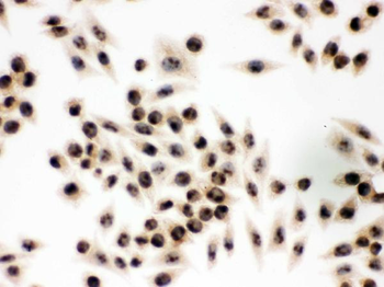
IHC analysis of EIF6 using anti-EIF6 antibody. EIF6 was detected in immunocytochemical section of SMMC-7721 cell. Heat mediated antigen retrieval was performed in citrate buffer (pH6, epitope retrieval solution) for 20 mins. The tissue section was blocked with 10% goat serum. The tissue section was then incubated with 1 µg/ml rabbit anti-EIF6 Antibody overnight at 4C. Biotinylated goat anti-rabbit IgG was used as secondary antibody and incubated for 30 minutes at 37C. The tissue section was developed using Strepavidin-Biotin-Complex (SABC) with DAB as the chromogen.
IHC analysis of EIF6 using anti-EIF6 antibody. EIF6 was detect
IHC analysis of EIF6 using anti-EIF6 antibody. EIF6 was detected in immunocytochemical section of SW480 cell. Enzyme antigen retrieval was performed using IHC enzyme antigen retrieval reagent for 15 mins. The cells were blocked with 10% goat serum. And then incubated with 1 µg/ml rabbit anti-EIF6 Antibody overnight at 4C. Biotinylated goat anti-rabbit IgG was used as secondary antibody and incubated for 30 minutes at 37C. The section was developed using Strepavidin-Biotin-Complex (SABC) with DAB as the chromogen.
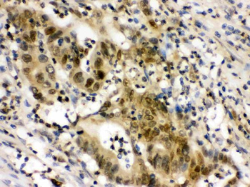
IHC analysis of EIF6 using anti-EIF6 antibody. EIF6 was detected in paraffin-embedded section of Human Intestinal Cancer Tissue. Heat mediated antigen retrieval was performed in citrate buffer (pH6, epitope retrieval solution) for 20 mins. The tissue section was blocked with 10% goat serum. The tissue section was then incubated with 1 µg/ml rabbit anti-EIF6 Antibody overnight at 4C. Biotinylated goat anti-rabbit IgG was used as secondary antibody and incubated for 30 minutes at 37C. The tissue section was developed using Strepavidin-Biotin-Complex (SABC) with DAB as the chromogen.
IHC analysis of EIF6 using anti-EIF6 antibody. EIF6 was detected in paraffin-embedded section of Mouse Intestine Tissue. Heat mediated antigen retrieval was performed in citrate buffer (pH6, epitope retrieval solution) for 20 mins. The tissue section was blocked with 10% goat serum. The tissue section was then incubated with 1 µg/ml rabbit anti-EIF6 Antibody overnight at 4C. Biotinylated goat anti-rabbit IgG was used as secondary antibody and incubated for 30 minutes at 37C. The tissue section was developed using Strepavidin-Biotin-Complex (SABC) with DAB as the chromogen.
IHC analysis of EIF6 using anti-EIF6 antibody. EIF6 was detected in paraffin-embedded section of Rat Testis Tissue. Heat mediated antigen retrieval was performed in citrate buffer (pH6, epitope retrieval solution) for 20 mins. The tissue section was blocked with 10% goat serum. The tissue section was then incubated with 1 µg/ml rabbit anti-EIF6 Antibody overnight at 4C. Biotinylated goat anti-rabbit IgG was used as secondary antibody and incubated for 30 minutes at 37C. The tissue section was developed using Strepavidin-Biotin-Complex (SABC) with DAB as the chromogen.
Western blot analysis of EIF6 using anti-EIF6 antibody. Electrophoresis was performed on a 5-20% SDS-PAGE gel at 70V (Stacking gel) / 90V (Resolving gel) for 2-3 hours. The sample well of each lane was loaded with 30 ug of sample under reducing conditions. Lane 1: human Hela whole cell lysates, Lane 2: human K562 whole cell lysates, Lane 3: human 293T whole cell lysates, Lane 4: human HepG2 whole cell lysates, Lane 5: rat liver tissue lysates, Lane 6: mouse liver tissue lysates, Lane 7: mouse RAW264.7 whole cell lysates. After electrophoresis, proteins were transferred to a nitrocellulose membrane at 150 mA for 50-90 minutes. Blocked the membrane with 5% non-fat milk/TBS for 1.5 hour at RT. The membrane was incubated with rabbit anti-EIF6 antigen affinity purified polyclonal antibody at 0.5 µg/mL overnight at 4C, then washed with TBS-0.1% Tween 3 times with 5 minutes each and probed with a goat anti-rabbit IgG-HRP secondary antibody at a dilution of 1:5000 for 1.5 hour at RT. The signal is developed using an Enhanced Chemiluminescent detection (ECL) kit with Tanon 5200 system. A specific band was detected for EIF6 at approximately 26 k
* Mehrwertsteuer und Versandkosten nicht enthalten. Irrtümer und Preisänderungen vorbehalten