Flow cytometric analysis of Rsc96 cell using LOX-1 antibody.
Immunohistochemical staining of human lung carcinoma tissue using LOX-1 antibody.
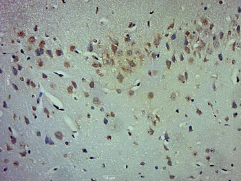
Paraformaldehyde-fixed, paraffin embedded (Mouse brain), Antigen retrieval by boiling in sodium citrate buffer (pH6.0) for 15 min, Block endogenous peroxidase by 3% hydrogen peroxide for 20 minutes, Blocking buffer (normal goat serum) at 37C for 30 min, Antibody incubation with (LOX 1) Polyclonal Antibody, Unconjugated (orb312297) at 1:500 overnight at 4C, followed by a conjugated secondary for 20 minutes and DAB staining.
Paraformaldehyde-fixed, paraffin embedded (Rabbit brain), Antigen retrieval by boiling in sodium citrate buffer (pH6.0) for 15 min, Block endogenous peroxidase by 3% hydrogen peroxide for 20 minutes, Blocking buffer (normal goat serum) at 37C for 30 min, Antibody incubation with (LOX 1) Polyclonal Antibody, Unconjugated (orb312297) at 1:400 overnight at 4C, followed by operating according to SP Kit (Rabbit) instructions and DAB staining.
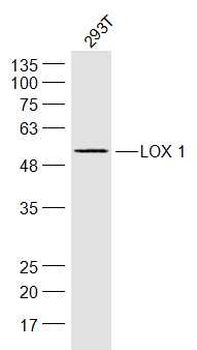
Sample: 293T (Human) Cell Lysate at 30 ug, Primary: Anti-LOX 1 (orb312297) at 1/300 dilution, Secondary: IRDye800CW Goat Anti-Rabbit IgG at 1/20000 dilution, Predicted band size: 31/50 kD, Observed band size: 50 kD.
Sample: Cerebrum (Rat) Lysate at 40 ug, Primary: Anti-LOX1 (orb312297) at 1/300 dilution, Secondary: IRDye800CW Goat Anti-Rabbit IgG at 1/20000 dilution, Predicted band size: 31/50 kD, Observed band size: 50 kD.
* Mehrwertsteuer und Versandkosten nicht enthalten. Irrtümer und Preisänderungen vorbehalten