

PPARGC1A Rabbit Polyclonal Antibody, Unconjugated
Artikelnummer:
BYT-ORB317701
- Bilder (5)


| Artikelname: | PPARGC1A Rabbit Polyclonal Antibody, Unconjugated |
| Artikelnummer: | BYT-ORB317701 |
| Hersteller Artikelnummer: | orb317701 |
| Alternativnummer: | BYT-ORB317701-50,BYT-ORB317701-100,BYT-ORB317701-200 |
| Hersteller: | Biorbyt |
| Wirt: | Rabbit |
| Kategorie: | Antikörper |
| Applikation: | ICC, IF, IHC-Fr, IHC-P, WB |
| Spezies Reaktivität: | Human |
| Immunogen: | KLH conjugated synthetic peptide derived from human PGC-1 (601-700/798aa) |
| Konjugation: | Unconjugated |
| Alternative Synonym: | AVPGC-1ALPHA, PPARGC-1A, LEM6, PGC-1(alpha), PGC-1alpha, PGC-1v, PGC1, PGC1A, PPARGC1, A830037N07Rik, Gm11133, PGC-1, PPARGC-1-alpha, Pgco1, LRPGC1, PGCvf, PGCvf-1, PGCvf1, PRGC1_BOVIN, PPARGC1A, PGC-1-alpha, PPAR-gamma coactivator 1-alpha, PRGC1_HUMAN, Ligand effect modulator 6, PRGC1_MOUSE, PRGC1_PIG, PRGC1_RAT, |
| PPARGC1A Rabbit Polyclonal Antibody |
| Klonalität: | Polyclonal |
| Konzentration: | 1mg/ml |
| Molekulargewicht: | 88 kDa |
| UniProt: | Q9UBK2 |
| Puffer: | 0.01M TBS (pH7.4) with 1% rAlbumin, 0.02% Proclin300 and 50% Glycerol. |
| Formulierung: | Liquid |
| Target-Kategorie: | PPARGC1A |
| Application Verdünnung: | WB=1:500-2000, IHC-P=1:100-500, IHC-F=1:100-500, ICC/IF=1:50-200, IF=1:100-500 |
|
|
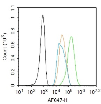
Blank control: HepG2. Primary Antibody (green line): Rabbit Anti-PPARGC1A antibody (orb317701), dilution: 1 µg/10 6 cells, Isotype Control Antibody (orange line): Rabbit IgG. Secondary Antibody: Goat anti-rabbit IgG-AF647, dilution: 1 µg/Test. Protocol, The cells were fixed with 4% PFA (10 min at room temperature) and then permeabilized with 90% ice-cold methanol for 20 min at -20C. The cells were then incubated in 5% BSA to block non-specific protein-protein interactions for 30 min at room temperature. Cells stained with Primary Antibody for 30 min at room temperature. The secondary antibody used for 40 min at room temperature. Acquisition of 20000 events was performed. |
|
|
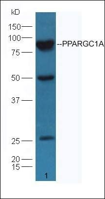
Sample: Lane 1: Recombinant human PPARGC1A, N-Trx-His, Primary: Anti-PPARGC1A (orb317701) at 1/1000 dilution, Secondary: IRDye800CW Goat Anti-Rabbit IgG at 1/20000 dilution, Predicted band size: 88 kDa, Observed band size: 61 kDa. |
|
|
Tissue/Cell: mouse liver tissue, 4% Paraformaldehyde-fixed and paraffin-embedded, Antigen retrieval: citrate buffer (0.01M, pH 6.0), Boiling bathing for 15 min, Block endogenous peroxidase by 3% Hydrogen peroxide for 30 min, Blocking buffer (normal goat serum) at 37C for 20 min, Incubation: Anti-PPARGC1A/PGC1 alpha Polyclonal Antibody, Unconjugated (orb317701) 1:200, overnight at 4C, followed by conjugation to the secondary antibody and DAB staining. |
|
|
Immunohistochemical staining of mouse kidney tissue using PPARGC1A antibody. |
|
|
Western blot analysis of mouse heart Cell lysates using PPARGC1A antibody. |
Produktgarantie und fachkundiger Support
