anti CD209 antibody, anti CD209 molecule antibody, anti CDSIGN antibody, anti CLEC4L antibody, anti DC-SIGN antibody, anti DC-SIGN1 antibody, anti C-type lectin domain family 4 member L antibody, anti C-type lectin domain family 4, member L antibody, anti CD209 antigen antibody, anti HIV gpl20-binding protein antibody, anti dendritic cell-specific ICAM-3-grabbing non-integrin 1 antibody, anti dendritic cell-spe antibody
Goat polyclonal antibody to CD209
Klonalität:
Polyclonal
Molekulargewicht:
45.8, 43.3, 45.0, 30.5, 41.0, 35.2, 27.4
Puffer:
Supplied at 0.5 mg/ml in Tris saline, 0.02% sodium azide, pH 7.3 with 0.5% bovine serum albumin. Aliquot and store at -20C. Minimize freezing and thawing.
Sequenz:
NRGEPNNVGEED
Target-Kategorie:
DC-SIGN / CD209
Application Verdünnung:
ELISA: 1:128000, WB: 0.1-0.3 µg/ml
Anwendungsbeschreibung:
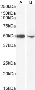
Application Notes: WB: Approx 50kDa band observed in Human Bone Marrow lysates (calculated MW of 45.8kDa according to NP_066978.1). The observed molecular weight corresponds to the glycosylated form. Recommended concentration: 0.1-0.3µg/ml
1 µg/ml staining of K562 (A) and (2 ug/ml) THP-1 (B) cell lysate (35 µg protein in RIPA buffer). Detected by chemiluminescence.
6 µg/ml staining of paraffin embedded Human Placenta. Heat induced antigen retrieval with citrate buffer pH 6, HRP-staining.
Negative Control showing staining of paraffin embedded Human Placenta, with no primary antibody.
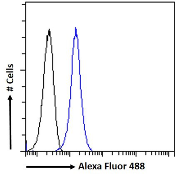
Flow cytometric analysis of paraformaldehyde fixed THP1 cells (blue line), permeabilized with 0.5% Triton. Primary incubation 1hr (10 ug/ml) followed by Alexa Fluor 488 secondary antibody (1 ug/ml). IgG control: Unimmunized goat IgG (black line) followed by Alexa Fluor 488 secondary antibody.
Immunofluorescence analysis of paraformaldehyde fixed THP-1 immobilized on Shi-fix(TM) plus cover-slips. Primary incubation 1hr (1:50 dilution) followed by Alexa Fluor 488 secondary antibody (1:2000 dilution), showing membrane and cytoplasmic staining. The nuclear stain is DAPI (blue). Negative control: Anti-Goat IgG followed by Alexa Fluor 488 secondary antibody.
* Mehrwertsteuer und Versandkosten nicht enthalten. Irrtümer und Preisänderungen vorbehalten