Anti-AKT3 Antibody was prepared from tissue culture supernatant by Protein A affinity chromatography using a synthetic peptide corresponding to internal residues of human AKT3 protein.
Preservative: 0.01% (w/v) Sodium Azide. Stabilizer: 10 mg/mL Bovine Serum Albumin (rAlbumin) - Immunoglobulin and Protease free, Buffer: 0.02 M Potassium Phosphate, 0.5 M Sodium Chloride, pH 7.2
Reinheit:
Anti-AKT3 antibody is directed against human AKT3. The antibody detects both unphosphorylated and phosphorylated forms of the protein. Anti-AKT3 antibody was purified from tissue culture by Protein A chromatography. Cross reactivity with AKT3 from other species has not been determined, however, the sequence of the immunogen shows 100% identity to human, mouse, and rat, therefore, cross reactivity is expected. Cross-reactivity with AKT2 and AKT has not been determined.
Formulierung:
Lyophilized
Application Verdünnung:
ELISA: User Optimized, FC: User Optimized, IHC: User Optimized, IF: User Optimized, WB: User Optimized
Anwendungsbeschreibung:
Application Notes: Anti-AKT3 FITC Antibody has been tested by ELISA and dot blot and is suitable for Flow Cytometry, immunohistochemistry, and western blotting. Expect a band approximately 56 kDa in size corresponding to AKT3 protein by western blotting in the appropriate cell lysate or extract. This monoclonal antibody reacts with human AKT. Specific conditions for reactivity should be optimized by the end user. For immunohistochemistry we recommend the use of fresh frozen tissues. Attempts at staining paraffin-embedded formalin fixed tissues were negative. No pre-treatment of sample is required
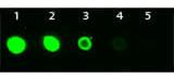
Dot Blot of Mouse anti-AKT3 Monoclonal Antibody Fluorescein Conjugated. Antigen: His-tagged AKT3. Load: Lane 1 - 100 ng Lane 2 - 33.3 ng Lane 3 - 11.1 ng Lane 4 - 3.70 ng Lane 5 - 1.23 ng. Primary antibody: n/a. Secondary antibody: Mouse anti-AKT3 Monoclonal Antibody Fluorescein Conjugated at 1:1000 for 60 min at RT. Block: orb348637 for 1 HR at RT.
Immunohistochemistry of Mouse Anti-AKT3 antibody. Tissue: human prostate carcinoma. A) AKT-3 antibody produced using CELLine, B) AKT-3 antibody produced using roller bottle. Fixation: formalin fixed paraffin embedded. Antigen retrieval: not required. Primary antibody: AKT-3 antibody at 10 µg/ml for 1 h at RT. Secondary antibody: Peroxidase mouse secondary antibody at 1:10000 for 1 h at RT. Localization: AKT3 is nuclear and occasionally cytoplasmic. Staining: AKT3 as precipitated brown signal with hematoxylin purple nuclear counterstain.
Immunohistochemistry of Mouse Monoclonal anti AKT3 Antibody in Mouse Embryonic Kidney. Tissue: Mouse Liver. Fixation: FFPE buffered formalin 10% conc. Ag Retrieval: Heat, Citrate pH6.2. Pressure Cooker. Primary antibody: anti-AKT3 at 2 ug/ml for 1.5 hour room Temp. Secondary Ab: MOUSE ON MOUSE HRP POLYMER 45 RT.
Western Blot of Mouse Anti-AKT3 antibody. Lane 1: C2C12. Lane 2: MEF1. Lane 3: MEF2. Lane 4: A549. Lane 5: Calu-1. Lane 6: PC3. Lane 7: HepG2. Lane 8: Jurkat. Lane 9: SKOV3. Lane 10: 293T. Load: 35 µg per lane. Primary antibody: AKT-3 antibody at 1:1000 for overnight at 4C. Secondary antibody: Anti mouse secondary antibody at 1:20000 for 1 h at RT. Block: 5% BLOTTO overnight at 4C. Predicted/Observed size: 56 kDa for AKT3.
Western Blot of Mouse anti-AKT3 antibody. Lane 1: Control. Lane 2: Rapa. Lane 3: T50. Lane 4: T250. Lane 5: Control. Lane 6: Rapa. Lane 7: T50. Lane 8: T250. Lane 9: AKT3 null. Load: 35 µg per lane. Primary antibody: AKT-3 antibody at 1:1000 for overnight at 4C. Secondary antibody: Anti mouse secondary antibody at 1:20000 for 1 h at RT. Block: 5% BLOTTO overnight at 4C. Predicted/Observed size: 56 kDa for AKT3.
Western Blot of Mouse anti-AKT3 antibody. Lane 1: GST Tagged recombinant AKT1. Lane 2: GST Tagged recombinant AKT2. Lane 3: GST Tagged recombinant AKT3. Load: 25 ng per lane. Primary antibody: AKT3 antibody at 1:1000 for overnight at 4C. Secondary antibody: Peroxidase mouse secondary antibody at 1:40000 for 30 min at RT. Block: orb348637 for 30 min at RT. Predicted/Observed size: 78 kDa for AKT3. Other band(s): none.
* Mehrwertsteuer und Versandkosten nicht enthalten. Irrtümer und Preisänderungen vorbehalten