Anti-SMAD3 pS423pS425 antibody was prepared from whole rabbit serum produced by repeated immunizations with a dual phosphorylated synthetic peptide corresponding to a c-terminal region with Serine 423 and Serine 425 of human SMAD3 protein.
Konjugation:
Unconjugated
Alternative Synonym:
rabbit anti-SMAD3 pS423pS425 antibody, SMAD-3, SMAD 3, mothers against decapentaplegic homolog 3 antibody, MAD homolog 3, Mothers against DPP homolog 3, SMAD family member 3, MADH3, MADH 3, JV15-2
Preservative: 0.01% (w/v) Sodium Azide. Stabilizer: None, Buffer: 0.02 M Potassium Phosphate, 0.15 M Sodium Chloride, pH 7.2
Reinheit:
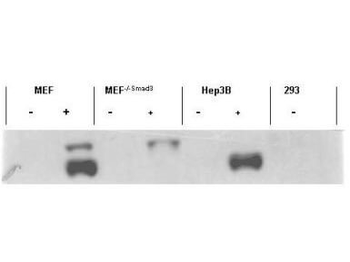
This affinity-purified antibody is directed against the phosphorylated form of human Smad3 protein at the pS423 and pS425 residues. The product was affinity purified from monospecific antiserum by immunoaffinity purification. Antiserum was first purified against the phosphorylated form of the immunizing peptide. The resultant affinity purified antibody was then cross adsorbed against the non-phosphorylated form of the immunizing peptide. Reactivity occurs against human Smad3 pS423 and pS425 protein and the antibody is specific for the phosphorylated form of the protein. Reactivity with non-phosphorylated human Smad3 is minimal by ELISA and western blot. Expect reactivity against phosphorylated Smad1 and Smad5. Negligible reactivity is seen against other phosphorylated Smad family members. A BLAST analysis was used to suggest cross reactivity with Smad3 from human, Xenopus laevis, Xenopus tropicalis, zebrafish, rat, mouse, swine, bovine and chicken based on 100% sequence homology with the immunogen. Reactivity against homologues from other sources is not known.
Application Notes: This affinity purified antibody has been tested for use in ELISA, immunohistochemistry and by western blot. Specific conditions for reactivity should be optimized by the end user. Expect a band approximately 48 kDa in size corresponding to phosphorylated Smad3 protein by western blotting in the appropriate stimulated tissue or cell lysate or extract. Less than 0.2% reactivity is observed against the non-phosphorylated form of the immunizing peptide. This antibody is phospho specific for dual phosphorylated pS423 and pS425 of Smad3. Stimulation with 2 ng/ml TGF-beta for 1 hour is suggested
AcSDKP suppresses TGFbeta/smad signaling and EndMT through the FGFR1/FRS2 pathway. (a) HMVECs were treated with N-FGFR1 for 48 h, and the FGFR1, TGFbetaR1 and TGFbetaR2 protein levels were analyzed by western blot. (b) HMVECs were treated with TGFbeta2 in the presence or absence of N-FGFR1 for 15 min with or without AcSDKP preincubation. The p-smad3 and TGFbetaR1 protein levels were analyzed by western blot. Densitometric analysis of the p-smad3/smad3 and TGFbetaR1/beta-actin levels (n = 3) in each group was performed. (c) HMVECs were incubated with either N-FGFR1 in the presence or absence of TGFbeta2 for 48 h with or without preincubation with AcSDKP for 2 h or with N-FGFR1 in the presence or absence of TGFbeta2 for 48 h with or without 24 h of incubation with FGF2 (50 ng/mL). The CD31, SM22alpha, FSP1 and alpha-SMA protein levels were analyzed by western blot. (d) HMVECs were transfected with FRS2 siRNA (100 nM) for 48 h with or without AcSDKP preincubation. The VE-cadherin, FSP1, vimentin, SM22alpha and p-smad3 levels were analyzed by western blot. (e) HMVECs were treated with N-FGFR1 for 48 h or 15 min in the presence or absence of N-TGFbeta (1, 2, 3) (1.0 µg/ml). The CD31, VE-cadherin, SM22alpha, FSP1, TGFbetaR1, TGFbetaR2 and p-smad3 levels were analyzed by western blot
MAP4K4 deficiency induces TGFbeta/smad signaling and EndMT via activation of integrin beta1. (a) HMVECs were transfected with MAP4K4 siRNA (100 nM) for 48 h. Next, the cells were treated with or without AcSDKP for 2 h. The p-smad3/smad3 pathway was analyzed by western blot. Densitometric analysis of the p-smad3/smad3 levels was performed, with n = 3 for each group. (b) HMVECs were treated with MAP4K4 siRNA for 48 h with or without AcSDKP treatment. The VE-cadherin, CD31, FSP1, SM22alpha and vimentin protein levels were analyzed by western blot. (c) HMVECs were transfected with MAP4K4 siRNA for 48 h in the presence or absence of TGFbeta2 with or without AcSDKP. The integrin beta1 level was analyzed by western blot.
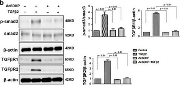
Proximity between AcSDKP and FGFR1 inhibits the TGFbeta/smad signaling pathway in HMVECs. (a) HMVECs were treated with N-FGFR1 (1.5 µg/ml) for 48 h with or without preincubation with AcSDKP (100 nM) for 2 h, and the proximity between AcSDKP and FGFR1 was analyzed by the Duolink In Situ Assay. For each slide, images at a * 400 original magnification were obtained from six different areas. (b and c) HMVECs were treated with TGFbeta2 (5 ng/mL) for 15 min or 48 h with or without preincubation with AcSDKP for 2 h, and the p-smad3, TGFbetaR1, TGFbetaR2 and FGFR1 levels were analyzed by western blot. Densitometric analysis of the p-smad3/smad3, TGFbetaR1/beta-actin, TGFbetaR2/beta-actin and FGFR1/beta-actin levels from each group (n = 6) were analyzed. (d and e) HMVECs were incubated with TGFbeta2 for 15 min or 48 h with or without preincubation with AcSDKP or its mutants (AcDSPK, AcSDKA, AcADKP) (100 nM) for 2 h. The p-smad3/smad3, TGFbetaR1/beta-actin, TGFbetaR2/beta-actin and FGFR1/beta-actin protein levels were analyzed by western blot.
Proximity between AcSDKP and FGFR1 inhibits the TGFbeta/smad signaling pathway in HMVECs. (a) HMVECs were treated with N-FGFR1 (1.5 µg/ml) for 48 h with or without preincubation with AcSDKP (100 nM) for 2 h, and the proximity between AcSDKP and FGFR1 was analyzed by the Duolink In Situ Assay. For each slide, images at a * 400 original magnification were obtained from six different areas. (b and c) HMVECs were treated with TGFbeta2 (5 ng/mL) for 15 min or 48 h with or without preincubation with AcSDKP for 2 h, and the p-smad3, TGFbetaR1, TGFbetaR2 and FGFR1 levels were analyzed by western blot. Densitometric analysis of the p-smad3/smad3, TGFbetaR1/beta-actin, TGFbetaR2/beta-actin and FGFR1/beta-actin levels from each group (n = 6) were analyzed. (d and e) HMVECs were incubated with TGFbeta2 for 15 min or 48 h with or without preincubation with AcSDKP or its mutants (AcDSPK, AcSDKA, AcADKP) (100 nM) for 2 h. The p-smad3/smad3, TGFbetaR1/beta-actin, TGFbetaR2/beta-actin and FGFR1/beta-actin protein levels were analyzed by western blot.
Biorbyts affinity purified anti-Sma
* Mehrwertsteuer und Versandkosten nicht enthalten. Irrtümer und Preisänderungen vorbehalten