CASZ1 Antibody, Unconjugated, Rabbit, Polyclonal Preis auf Anfrage
Artikelnummer:
BYT-ORB345676
Hersteller Artikelnummer:
orb345676
Alternativnummer:
BYT-ORB345676-25
Hersteller:
Biorbyt
Wirt:
Rabbit
Kategorie:
Antikörper
Applikation:
ELISA, WB
Spezies Reaktivität:
Human
Immunogen:
This affinity purified antibody was prepared from whole rabbit serum produced by repeated immunizations with a synthetic peptide corresponding to an internal region of Human Casz1 protein.
Konjugation:
Unconjugated
Alternative Synonym:
rabbit anti-CASZ1 Antibody, CASZ1, Zinc finger protein castor homolog 1, Castor-related protein, Zinc finger protein 693, CST, SRG, ZNF693
Preservative: 0.01% (w/v) Sodium Azide. Stabilizer: None, Buffer: 0.02 M Potassium Phosphate, 0.15 M Sodium Chloride, pH 7.2
Reinheit:
This affinity purified antibody is directed against human Casz1 protein. The product was affinity purified from monospecific antiserum by immunoaffinity purification. A BLAST analysis was used to suggest reactivity with CASZ1 proteins from human, mouse, Drosophila, chimpanzee, and macaque based on a 100% homology. Partial reactivity is expected with horse and dog CASZ1 based on a 92% homology with the immunizing sequence. Cross-reactivity with CASZ1 from other sources has not been determined.
Formulierung:
Liquid (sterile filtered)
Application Verdünnung:
ELISA: 1:45,000, WB: 1:1,000 - 1:5,000
Anwendungsbeschreibung:
Application Notes: This antibody has been tested for use in ELISA, IHC, IF, and western blotting. This antiserum detects endogenous Casz1 proteins, both the hCasz5 and hCasz11 isoforms. Expect a band approximately 125 kDa for hCasz5 and 190 kDa for hCaz11 in size corresponding Casz1 by western blotting in the appropriate cell lysate or extract. Specific conditions for reactivity should be optimized by the end user
Immunofluorescence of Rabbit anti-CASZ1 Antibody. Tissue: adult murine ocular tissue. Antibody: Rabbit Anti-CASZ1 Antibody. Counterstain: DAPI. Localization: nucleus in lens epithelia but primarily localizes in the cytoplasm in photoreceptor cells.
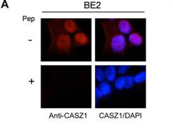
Immunofluorescence results of Endogenous CASZ1. Cells: BE2 cells. With or without Pre-Incubation of Anti-CASZ1 Antibody with CASZ1 Peptide. Staining: Rabbit Anti-CASZ1 Antibody. Chromatin counter stain: DAPI.
Immunofluorescence results of Rabbit Anti-CASZ1 Antibody. Tissue: Mouse Xenograft tumor of human NB cell line transfected with or without tetracycline inducible CASZ1 (NGPtetCASZ1a). Antibody: Rabbit Anti-CASZ1 Antibody. Counterstain: DAPI.
Immunohistochemistry results of Rabbit Anti-hCasz1 Antibody. Tissue: NB patient tumor. A. CASZ1 localized exclusively in the cytoplasm. B. CASZ1 localized in the cytoplasm and nucleus. Primary Antibody: Rabbit Anti-CASZ1 stained brown. Nucleus counterstained with hematoxylin (blue).
Immunohistochemistry results of Rabbit Anti-hCASZ1 Antibody. Tissue: NB patient tumor. A. Score 0- a rare positive nuclei. B. Score 1- (1-10% positive) equivocal/uninterpretable. C. Score 2- (10-50% positive) weak positive. D. Score 3- (> 50% positive) strong positive. Primary Antibody: Rabbit Anti-CASZ1 stained brown. Nucleus counterstained with hematoxylin (blue). Localization: Nuclear.
Western Blot of Anti-CASZ1 Antibody. Lane 1: NBLS Cytoplasmic (20 µg). Lane 2: NBLS Nuclear (3 µg). Lane 3: BE2C Cytoplasmic (30 µg). Lane 4: BE2C Nuclear (7 µg). Lane 5: SY5Y-CASZ1b (10 µg). Block: 5% Blotto/TTBS for 1 hour. Primary: Casz1 1:10000 for 1 hour. Secondary: Goat anti-Rabbit HRP for 1 hour. 240sec exposure. Detects nuclear endogenous CASZ1a and CASZ1b, and transiently transfected CASZ1b isoform.
Western blot using Biorbyts anti-hCASZ1 antibody. This blot shows detection of endogenous and transfected human CASZ1 protein in fresh whole cell lysate (~30 µg). Lane 1: BE2(s) cell lysate. Lane 2: BE2(N) cell lysate. Lane 3: SY5Y transfected with hCASZ5 (125kDa). Lane 4: SY5Y transfected with hCASZ11 (190kDa). Protein was resolved by SDS-PAGE and transferred onto nitrocellulose. After blocking, the membrane was probed with the primary antibody diluted to 1:1000 for 1.5 hours at room temperature then incubated with HRP-conjugated Goat Anti-Rabbit antibody for 45 min. at room temperature.
* Mehrwertsteuer und Versandkosten nicht enthalten. Irrtümer und Preisänderungen vorbehalten