Rabbit IgG whole molecule Texas Red conjugate, Rabbit IgG Texas Red conjugation
Rabbit IgG Texas Red Antibody
Konzentration:
1.0 mg/mL
Puffer:
Preservative: 0.01% (w/v) Sodium Azide. Stabilizer: 10 mg/mL Bovine Serum Albumin (rAlbumin) - Immunoglobulin and Protease free, Buffer: 0.02 M Potassium Phosphate, 0.15 M Sodium Chloride, pH 7.2
Quelle:
Rabbit
Reinheit:
This product was prepared from normal serum by delipidation, salt fractionation, ion exchange chromatography followed by extensive dialysis against the buffer stated above. Assay by immunoelectro-phoresis resulted in a single precipitin arc against anti-Rabbit IgG and anti-Rabbit Serum.
Formulierung:
Lyophilized
Anwendungsbeschreibung:
Biological Origin: Rabbit. Application Notes: Rabbit IgG whole molecule Texas Red is designed for immunofluorescence microscopy, fluorescence based plate assays (FLISA) and fluorescent western blotting. This product is also suitable for multiplex analysis, including multicolor imaging, utilizing various commercial platforms
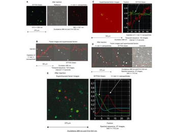
Case of temporal and spectral observations of iron nanoparticles conjugated with Texas Red injected in a culture of untreated murine cardiac HL1-NB cells counterstained with SYTOX Green in which emissions are collected through band-pass filters. Goat anti-rabbit IgG microbeads were incubated with Texas Red conjugated rabbit IgG (p/n orb346327) in order to obtain fluorescent nanoparticles. A) Regular mode through band-pass filters after injection. B) The emission is then collected in the temporal mode in a long-pass-filter and processed by means of FAMIS. A stable emission corresponding to SYTOX Green and an emission uptake corresponding to red nanoparticles are visualized to localize nanoparticles in cell compartments. C) Superimposition in true color of the factor image. In some cells, the presence of a high signal emphasizes the fact that nanoparticles accumulate inside cytoplasm. D) Spectral mode through 10 nm band-pass filters. Investigation by means of FAMIS. A green emission (535 nm) corresponding to SYTOX Green in the first factor image and a red emission (610 nm) corresponding to red nanoparticles are visualized in the second factor image. Nanoparticles are either captured or not by the cells. E) Superimposition in true color of these factor images is performed to localize nanoparticles in cell compartments.
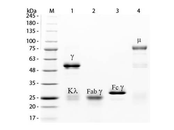
SDS-PAGE of Rabbit IgG Whole Molecule Rhodamine Conjugated (p/n orb346302). Lane M: 3 µl Opal Prestained Marker. Lane 1: Reduced Rabbit IgG Whole Molecule Rhodamine Conjugated (p/n orb346302). Lane 2: Reduced Rabbit IgG F(ab) Fragment (p/n orb2652745). Lane 3: Reduced Rabbit IgG F(c) Fragment (p/n orb346310). Lane 4: Reduced Rabbit IgM Whole Molecule (p/n orb346313). Load: 1 µg for F(ab) and F(c), 1.2 µg for IgG and IgM. Predicted/Observed size: IgG at 50 and 25 kDa, F(c) at 25 kDa, F(ab) at 25 kDa, IgM at 70 and 23 kDa. Observed F(c) Fragment migrates slightly higher.
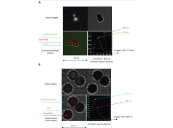
Spectral observations of FTR (FITC + Texas Red) beads in which (A) emission and (B) excitation sequences are collected and processed by FAMIS. Texas Red-conjugated rabbit IgG (p/n orb346327). Abbreviation: FAMIS, factor analysis of medical image sequences.
* Mehrwertsteuer und Versandkosten nicht enthalten. Irrtümer und Preisänderungen vorbehalten