IF image of rat thymus tissue using CD52 antibody (primary antibody at 5 ug/ml)
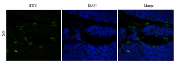
Immunofluorescence staining of fixed rat thymus tissue with anti-CD52 antibody Campath-1H. Immunofluorescence analysis of fixed rat thymus tissue stained with the chimeric human IgG1 version of Campath-1H (orb348863) at 5 µg/ml followed by a FITC conjugated secondary antibody, showing membrane and cytoplasmic staining of a subset of cells. The nuclear stain is DAPI (blue). Panels show from left-right, orb348863 (FITC), DAPI and merged channels.
Immunofluorescent staining of rat thymus using anti-CD52 antibody. Formaldehyde-fixed rat thymus slices were stained with orb348863 at 5 µg/ml and detected with a FITC-conjugated secondary antibody.
Immunohistochemical staining of human thyroid cancer using anti-CD52 antibody. Formalin fixed human thyroid cancer slices were were stained with a orb348863 at 5 µg/ml.
Immunohistochemical staining of rat epididymis tissue using anti-CD52 antibody Campath-1H. Anti-CD52 staining of formaldehyde fixed paraffin embedded rat epididymis tissue, at 40x magnification. The human IgG1-chimeric version of Campath-1H (orb348863) was used to stain samples at a concentration of 5 µg/ml.
Immunohistochemical staining of rat spleen tissue using anti-CD52 antibody Campath-1H. Anti-CD52 staining of formaldehyde fixed paraffin embedded rat spleen tissue, at 40x magnification. The human IgG1-chimeric version of Campath-1H (orb348863) was used to stain samples at a concentration of 5 µg/ml.
Immunohistochemical staining of rat thymus tissue using anti-CD52 antibody Campath-1H. Anti-CD52 staining of formaldehyde fixed paraffin embedded rat thymus tissue, at 40x magnification. The human IgG1-chimeric version of Campath-1H (orb348863) was used to stain samples at a concentration of 5 µg/ml.
* Mehrwertsteuer und Versandkosten nicht enthalten. Irrtümer und Preisänderungen vorbehalten