E. coli-derived human ADK recombinant protein (Position: K165-T351). Human ADK shares 88.8% and 88.2% amino acid (aa) sequence identity with mouse and rat ADK, respectively.
Each vial contains 4 mg Trehalose, 0.9 mg NaCl and 0.2 mg Na2HPO4.
Formulierung:
Lyophilized
Target-Kategorie:
Adenosine kinase
Application Verdünnung:
Western blot, 0.1-0.5µg/ml, Human, Mouse, Rat Immunohistochemistry (Paraffin-embedded Section), 2-5µg/ml, Human, Mouse, Rat Immunocytochemistry/Immunofluorescence, 5µg/ml, Human
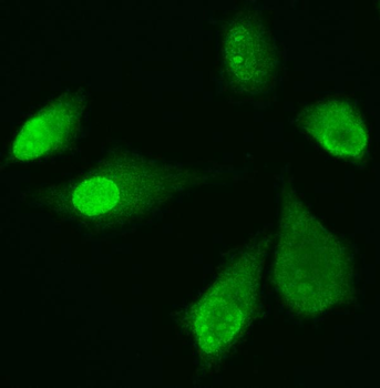
IF analysis of ADK using anti-ADK antibody. ADK was detected in an immunocytochemical section of PC-3 cells. Enzyme antigen retrieval was performed using IHC enzyme antigen retrieval reagent for 15 mins. The cells were blocked with 10% goat serum. And then incubated with 5 µg/mL rabbit anti-ADK Antibody overnight at 4C. DyLight488 Conjugated Goat Anti-Rabbit IgG was used as secondary antibody at 1:100 dilution and incubated for 30 minutes at 37C. Visualize using a fluorescence microscope and filter sets appropriate for the label used.
WB analysis of ADK using anti-ADK antibody.Lane 1:rat liver tissue, 2:rat kidney tiss
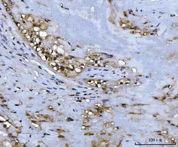
IHC analysis of ADK using anti-ADK antibody. ADK was detected in a paraffin-embedded section of human placenta tissue. Heat mediated antigen retrieval was performed in EDTA buffer (pH8.0, epitope retrieval solution). The tissue section was blocked with 10% goat serum. The tissue section was then incubated with 2 µg/ml rabbit anti-ADK Antibody overnight at 4C. Peroxidase Conjugated Goat Anti-rabbit IgG was used as secondary antibody and incubated for 30 minutes at 37C. The tissue section was developed using HRP Conjugated Rabbit IgG Super Vision Assay Kit with DAB as the chromogen.
IHC analysis of ADK using anti-ADK antibody. ADK was detected in a paraffin-embedded section of mouse liver tissue. Heat mediated antigen retrieval was performed in EDTA buffer (pH8.0, epitope retrieval solution). The tissue section was blocked with 10% goat serum. The tissue section was then incubated with 2 µg/ml rabbit anti-ADK Antibody overnight at 4C. Peroxidase Conjugated Goat Anti-rabbit IgG was used as secondary antibody and incubated for 30 minutes at 37C. The tissue section was developed using HRP Conjugated Rabbit IgG Super Vision Assay Kit with DAB as the chromogen.
IHC analysis of ADK using anti-ADK antibody. ADK was detected in a paraffin-embedded section of rat liver tissue. Heat mediated antigen retrieval was performed in EDTA buffer (pH8.0, epitope retrieval solution). The tissue section was blocked with 10% goat serum. The tissue section was then incubated with 2 µg/ml rabbit anti-ADK Antibody overnight at 4C. Peroxidase Conjugated Goat Anti-rabbit IgG was used as secondary antibody and incubated for 30 minutes at 37C. The tissue section was developed using HRP Conjugated Rabbit IgG Super Vision Assay Kit with DAB as the chromogen.
Western blot analysis of ADK using anti-ADK antibody. Electrophoresis was performed on a 5-20% SDS-PAGE gel at 70V (Stacking gel) / 90V (Resolving gel) for 2-3 hours. The sample well of each lane was loaded with 30 ug of sample under reducing conditions. Lane 1: human placenta tissue lysates, Lane 2: human PC-3 whole cell lysates, Lane 3: human T47D whole cell lysates, Lane 4: human U937 whole cell lysates, Lane 5: human U251 whole cell lysates, Lane 6: human HepG2 whole cell lysates. red to a nitrocellulose membrane at 150 mA for 50-90 minutes. Blocked the membrane with 5% non-fat milk/TBS for 1.5 hour at RT. The membrane was incubated with rabbit anti-ADK antigen affinity purified polyclonal antibody at 0.5 µg/mL overnight at 4C, then washed with TBS-0.1% Tween 3 times with 5 minutes each and probed with a goat anti-rabbit IgG-HRP secondary antibody at a dilution of 1:5000 for 1.5 hour at RT. The signal is developed using an Enhanced Chemiluminescent detection (ECL) kit with Tanon 5200 system. A specific band was detected for ADK at approximately 45 kDa. The expected band size for ADK is at 45 kDa.
Western blot analysis of ADK using anti-ADK antibody. Electrophoresis was performed on a 5-20% SDS-PAGE gel at 70V (Stacking gel) / 90V (Resolving gel) for 2-3 hours. The sample well of each lane was loaded with 30 ug of sample under reducing conditions. Lane 1: rat liver tissue lysates, Lane 2: rat kidney tissue lysates, Lane 3: rat stomach tissue lysates, Lane 4: rat PC-12 whole cell lysates, Lane 5: mouse kidney tissue lysates, Lane 6: mouse stomach tissue lysates, Lane 7: mouse NIH/3T3 whole cell lysates. red to a nitrocellulose membrane at 150 mA for 50-90 minute
* Mehrwertsteuer und Versandkosten nicht enthalten. Irrtümer und Preisänderungen vorbehalten