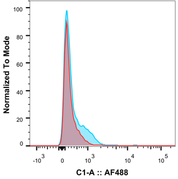
Flow-cytometry using anti-CD3 antibody YTH 12.5 Rhesus monkey lymphocytes were stained with an isotype control (orb256390, red) or the rabbit-chimeric version of YTH 12.5 (orb411552, blue) at a concentration of 1 µg/ml for 30 mins at RT. After washing, bound antibody was detected using a AF488 conjugated donkey anti-rabbit antibody and cells analysed on a FlowJo single-cell flow cytometer.
Flow-cytometry using anti-CD3 epsilon antibody YTH 12.5 Human lymphocytes were stained with an isotype control (orb256390, panel A) or the rabbit-chimeric version of YTH 12.5 (orb411552, panel B) at a concentration of 1 µg/ml for 30 mins at RT. After washing, bound antibody was detected using a AF488 conjugated donkey anti-rabbit antibody and cells analysed on a FACSCanto flow-cytometer.
Flow-cytometry using the anti-CD3E antibody YTH 12.5 Jurkat cells were stained with unimmunized rabbit IgG antibody (black line) or the rabbit-chimeric version of YTH 12.5 (orb411552, blue line) at a concentration of 10 µg/ml for 30 mins at RT. After washing, bound antibody was detected using anti-rabbit IgG JK (FITC-conjugate) antibody (129936) at 2 µg/ml and cells analyzed on a FACSCanto flow-cytometer.
Immunohistochemical staining of human tonsil tissue using anti-CD3E antibody YTH 12.5 Anti-CD3E staining of paraffin embedded human tissue using the rabbit-chimeric version of YTH 12.5 (orb411552). Antigen retreival was acheived by microwaving in citrate buffer (pH6), followed by blocking with protein block serum-free buffer. Primary antibody incubation with orb411552 was carried out at 4 µg/ml for 30 minutes. Samples were then incubated with an anti-rabbit IgG HRP secondary antibody for 20 mins followed by DAB (3, 3'-diaminobenzidine), and counter-staining with haemotoxylin. Strong membrane staining of T-cells at the periphery of the germinal centre and between follicles may be observed. Recommended concentration, 2-4 µg/ml.
Western Blot using anti-CD3E antibody YTH 12.5 Human spleen sample (35 µg protein in RIPA buffer) were resolved on a 10% SDS PAGE gel and blots probed with the chimeric rabbit version of YTH 12.5 (orb411552) at 0.3 µg/ml before detection using an anti-rabbit secondary antibody. A primary incubation of 1h was used and protein was detected by chemiluminescence. The expected band size for CD3E is 23.1 kDa. orb411552 successfully detected human CD3E.
* Mehrwertsteuer und Versandkosten nicht enthalten. Irrtümer und Preisänderungen vorbehalten