E. coli-derived human RUNX1T1/ETO recombinant protein (Position: T335-D510).
Konjugation:
Unconjugated
Alternative Synonym:
AML1T1, CBFA2T1, CDR, Cyclin D related protein, Eight twenty one protein, ETO, MTG8, MTG8b, Protein CBFA2T1, Protein ETO, Protein MTG8, RUNX1T1, ZMYND2
Anti-RUNX1T1/ETO Antibody. Tested in ELISA, Flow Cytometry, IF, IHC, IHC-F, ICC, WB applications. This antibody reacts with Human, Mouse, Rat.
Klonalität:
Polyclonal
Konzentration:
Adding 0.2 ml of distilled water will yield a concentration of 500 µg/ml.
Western blot, 0.1-0.5µg/ml, Human, Mouse Immunohistochemistry (Paraffin-embedded Section), 0.5-1µg/ml, Human, Mouse Immunocytochemistry/Immunofluorescence, 2µg/ml, Human Immunoprecipitation, 0.5-2 µg/ml, Human Flow Cytometry (Fixed), 1-3µg/1x10 6 cells, H
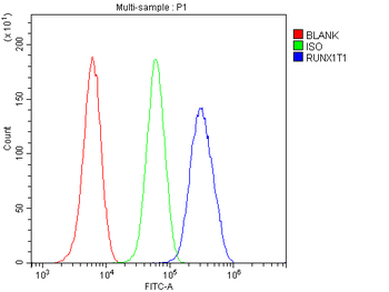
Flow Cytometry analysis of U20S cells using anti-RUNX1T1 antibody. Overlay histogram showing U20S cells (Blue line). To facilitate intracellular staining, cells were fixed with 4% paraformaldehyde and permeabilized with permeabilization buffer. The cells were blocked with 10% normal goat serum. And then incubated with rabbit anti-RUNX1T1 Antibody (1 µg/1x10 6 cells) for 30 min at 20C. DyLight488 conjugated goat anti-rabbit IgG (5-10 µg/1x10 6 cells) was used as secondary antibody for 30 minutes at 20C. Isotype control antibody (Green line) was rabbit IgG (1 µg/1x10 6) used under the same conditions. Unlabelled sample (Red line) was also used as a control.
WB analysis of RUNX1T1/ETO using anti-RUNX1T1/ETO antibody.Lane 1
IF analysis of RUNX1T1 using anti-RUNX1T1 antibody. RUNX1T1 was detected in immunocytochemical section of U20S cell. Enzyme antigen retrieval was performed using IHC enzyme antigen retrieval reagent for 15 mins. The cells were blocked with 10% goat serum. And then incubated with 2 µg/mL rabbit anti-RUNX1T1 Antibody overnight at 4C. DyLight488 Conjugated Goat Anti-Rabbit IgG was used as secondary antibody at 1:100 dilution and incubated for 30 minutes at 37C. Visualize using a fluorescence microscope and filter sets appropriate for the label used.
IHC analysis of RUNX1T1/ETO using anti-RUNX1T1/ETO antibody. RUNX1T1/ETO was detected in paraffin-embedded section of human mammary cancer tissue. Heat mediated antigen retrieval was performed in citrate buffer (pH6, epitope retrieval solution) for 20 mins. The tissue section was blocked with 10% goat serum. The tissue section was then incubated with 1 µg/ml rabbit anti-RUNX1T1/ETO Antibody overnight at 4C. Biotinylated goat anti-rabbit IgG was used as secondary antibody and incubated for 30 minutes at 37C. The tissue section was developed using Strepavidin-Biotin-Complex (SABC) with DAB as the chromogen.
IHC analysis of RUNX1T1/ETO using anti-RUNX1T1/ETO antibody. RUNX1T1/ETO was detected in paraffin-embedded section of mouse brain tissue. Heat mediated antigen retrieval was performed in citrate buffer (pH6, epitope retrieval solution) for 20 mins. The tissue section was blocked with 10% goat serum. The tissue section was then incubated with 1 µg/ml rabbit anti-RUNX1T1/ETO Antibody overnight at 4C. Biotinylated goat anti-rabbit IgG was used as secondary antibody and incubated for 30 minutes at 37C. The tissue section was developed using Strepavidin-Biotin-Complex (SABC) with DAB as the chromogen.
Western blot analysis of RUNX1T1/ETO using anti-RUNX1T1/ETO antibody. Electrophoresis was performed on a 5-20% SDS-PAGE gel at 70V (Stacking gel) / 90V (Resolving gel) for 2-3 hours. The sample well of each lane was loaded with 50 ug of sample under reducing conditions. Lane 1: human placenta tissue lysates, Lane 2: mouse testis tissue lysates. After Electrophoresis, proteins were transferred to a Nitrocellulose membrane at 150mA for 50-90 minutes. Blocked the membrane with 5% Non-fat Milk/ TBS for 1.5 hour at RT. The membrane was incubated with rabbit anti-RUNX1T1/ETO antigen affinity purified polyclonal antibody at 0.5 µg/mL overnight at 4C, then washed with TBS-0.1% Tween 3 times with 5 minutes each and probed with a goat anti-rabbit IgG-HRP secondary antibody at a dilution of 1:10000 for 1.5 hour at RT. The signal is developed using an Enhanced Chemiluminescent detection (ECL) kit with Tanon 5200 system. A specific band was detected for RUNX1T1/ETO at approximately 67KD. The expected band size for RUNX1T1/ETO is at 67KD.
* Mehrwertsteuer und Versandkosten nicht enthalten. Irrtümer und Preisänderungen vorbehalten