
DIO2 Rabbit Polyclonal Antibody, Unconjugated
Artikelnummer:
BYT-ORB5972
- Bilder (8)
| Artikelname: | DIO2 Rabbit Polyclonal Antibody, Unconjugated |
| Artikelnummer: | BYT-ORB5972 |
| Hersteller Artikelnummer: | orb5972 |
| Alternativnummer: | BYT-ORB5972-50,BYT-ORB5972-100,BYT-ORB5972-200 |
| Hersteller: | Biorbyt |
| Wirt: | Rabbit |
| Kategorie: | Antikörper |
| Applikation: | IF, IHC-Fr, IHC-P, WB |
| Spezies Reaktivität: | Human, Mouse, Rat |
| Immunogen: | KLH conjugated synthetic peptide derived from human DIO2 (101-200/273aa) |
| Konjugation: | Unconjugated |
| Alternative Synonym: | 5DII, DIOII, D2, SELENOY, SelY, TXDI2, IOD2_BOVIN, DIO2, Type 2 DI, Type-II 5-deiodinase, 1.21.99.4, IOD2_HUMAN, ITDI2, IOD2_MOUSE, IOD2_PIG, IOD2_RAT, |
| DIO2 Rabbit Polyclonal Antibody |
| Klonalität: | Polyclonal |
| Konzentration: | 1mg/ml |
| Molekulargewicht: | 30 kDa |
| UniProt: | Q92813 |
| Puffer: | 0.01M TBS (pH7.4) with 1% rAlbumin, 0.02% Proclin300 and 50% Glycerol. |
| Formulierung: | Liquid |
| Target-Kategorie: | DIO2 |
| Application Verdünnung: | WB=1:500-2000, IHC-P=1:100-500, IHC-F=1:100-500, IF=1:100-500 |
|
|
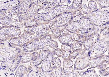
Paraformaldehyde-fixed, paraffin embedded (human placenta), Antigen retrieval by boiling in sodium citrate buffer (pH6.0) for 15 min, Block endogenous peroxidase by 3% hydrogen peroxide for 20 minutes, Blocking buffer (normal goat serum) at 37C for 30 min, Incubation with (DIO2) Polyclonal Antibody, Unconjugated (orb5972) at 1:200 overnight at 4C, followed by operating according to SP Kit (Rabbit) instructionsand DAB staining. |
|
|
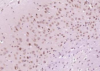
Paraformaldehyde-fixed, paraffin embedded (Mouse brain), Antigen retrieval by boiling in sodium citrate buffer (pH6.0) for 15 min, Block endogenous peroxidase by 3% hydrogen peroxide for 20 minutes, Blocking buffer (normal goat serum) at 37C for 30 min, Antibody incubation with (DIO2) Polyclonal Antibody, Unconjugated (orb5972) at 1:400 overnight at 4C, followed by operating according to SP Kit (Rabbit) instructionsand DAB staining. |
|
|
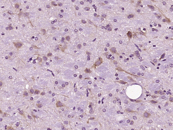
Paraformaldehyde-fixed, paraffin embedded (Rat brain), Antigen retrieval by boiling in sodium citrate buffer (pH6.0) for 15 min, Block endogenous peroxidase by 3% hydrogen peroxide for 20 minutes, Blocking buffer (normal goat serum) at 37C for 30 min, Antibody incubation with (DIO2) Polyclonal Antibody, Unconjugated (orb5972) at 1:400 overnight at 4C, followed by operating according to SP Kit (Rabbit) instructions and DAB staining. |
|
|
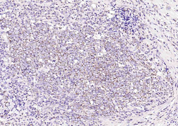
Paraformaldehyde-fixed, paraffin embedded (Thyroid tumor), Antigen retrieval by boiling in sodium citrate buffer (pH6.0) for 15 min, Block endogenous peroxidase by 3% hydrogen peroxide for 20 minutes, Blocking buffer (normal goat serum) at 37C for 30 min, Incubation with (DIO2) Polyclonal Antibody, Unconjugated (orb5972) at 1:200 overnight at 4C, followed by operating according to SP Kit (Rabbit) instructionsand DAB staining. |
|
|
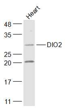
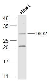
Sample: Heart (Mouse) Lysate at 40 ug, Primary: Anti-DIO2 (orb5972) at 1/1000 dilution, Secondary: IRDye800CW Goat Anti-Rabbit IgG at 1/20000 dilution, Predicted band size: 30 kD, Observed band size: 30 kD. |
|
|
WB analysis of mouse heart Lysate at 40 ug using DIO2 antibody |
|
|
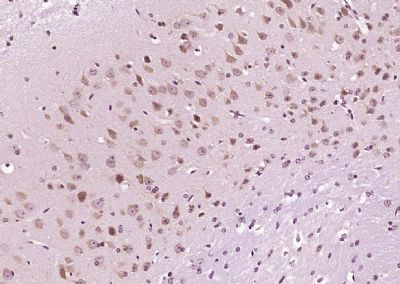
IHC-P image of Mouse brain tissue using DIO2 antibody |
|
|
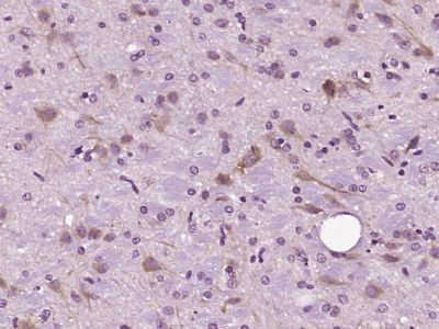
IHC-P image of rat brain tissue using DIO2 antibody |
Produktgarantie und fachkundiger Support
