Western blot, 0.1-0.5µg/ml, Human, Mouse, Rat Immunocytochemistry/Immunofluorescence,2µg/ml,Human Flow Cytometry (Fixed), 1-3µg/1x10 6 cells, Human
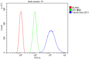
Flow Cytometry analysis of HEPA1-6 cells using anti-Tubulin beta antibody. Overlay histogram showing HEPA1-6 cells (Blue line). To facilitate intracellular staining, cells were fixed with 4% paraformaldehyde and permeabilized with permeabilization buffer. The cells were blocked with 10% normal goat serum. And then incubated with mouse anti-Tubulin beta Antibody (1 µg/1x10 6 cells) for 30 min at 20C. DyLight488 conjugated goat anti-mouse IgG (5-10 µg/1x10 6 cells) was used as secondary antibody for 30 minutes at 20C. Isotype control antibody (Green line) was mouse IgG (1 µg/1x10 6) used under the same conditions. Unlabelled sample without incubation with primary antibody and secondary antibody (Red line) was used as a blank control.
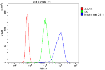
Flow Cytometry analysis of U937 cells using anti-Tubulin beta antibody. Overlay histogram showing U937 cells (Blue line). To facilitate intracellular staining, cells were fixed with 4% paraformaldehyde and permeabilized with permeabilization buffer. The cells were blocked with 10% normal goat serum. And then incubated with mouse anti-Tubulin beta Antibody (1 µg/1x10 6 cells) for 30 min at 20C. DyLight488 conjugated goat anti-mouse IgG (5-10 µg/1x10 6 cells) was used as secondary antibody for 30 minutes at 20C. Isotype control antibody (Green line) was mouse IgG (1 µg/1x10 6) used under the same conditions. Unlabelled sample without incubation with primary antibody and secondary antibody (Red line) was used as a blank control.
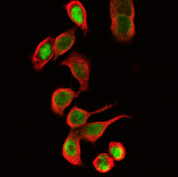
IF analysis of Tubulin beta using anti-Tubulin beta antibody and anti-FOSB antibody. Tubulin beta was detected in immunocytochemical section of CACO-2 cells. Enzyme antigen retrieval was performed using IHC enzyme antigen retrieval reagent for 15 mins. The cells were blocked with 10% goat serum. And then incubated with 2 µg/mL mouse anti-Tubulin beta Antibody and rabbit anti-FOSB antibody overnight at 4C. DyLight594 Conjugated Goat Anti-Mouse IgG and DyLight488 Conjugated Goat Anti-Rabbit IgG were used as secondary antibody at 1:100 dilution and incubated for 30 minutes at 37C. Visualize using a fluorescence microscope and filter sets appropriate for the label used.
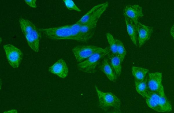
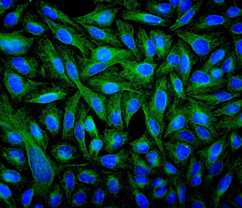
IF analysis of Tubulin beta using anti-Tubulin beta antibody. Tubulin beta was detected in immunocytochemical section of U2OS cell. Enzyme antigen retrieval was performed using IHC enzyme antigen retrieval reagent for 15 mins. The cells were blocked with 10% goat serum. And then incubated with 2 µg/mL mouse anti-Tubulin beta Antibody overnight at 4C. DyLight488 Conjugated Goat Anti-Mouse IgG was used as secondary antibody at 1:100 dilution and incubated for 30 minutes at 37C. The section was counterstained with DAPI. Visualize using a fluorescence microscope and filter sets appropriate for the label used.
IF analysis of Tubulin beta using anti-Tubulin beta antibody. Tubulin beta was detected in immunocytochemical section of U2OS cell. Enzyme antigen retrieval was performed using IHC enzyme antigen retrieval reagent for 15 mins. The cells were blocked with 10% goat serum. And then incubated with 2 µg/mL mouse anti-Tubulin beta Antibody overnight at 4C. DyLight488 Conjugated Goat Anti-Mouse IgG was used as secondary antibody at 1:100 dilution and incubated for 30 minutes at 37C. The section was counterstained with DAPI. Visualize using a fluorescence microscope and filter sets appropriate for the label used.
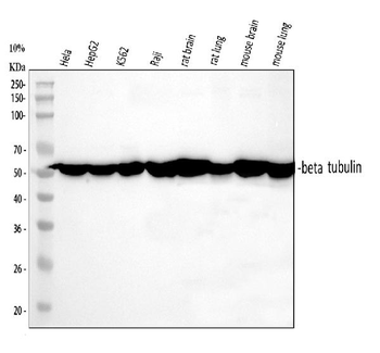
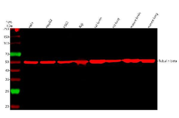
Western blot analysis of Tubulin beta using anti-Tubulin beta antibody. Electrophoresis was performed on a 5-20% SDS-PAGE gel at 70V (Stacking gel) / 90V (Resolving gel) for 2-3 hours. The sample well of each lane was loaded with 30 ug of sample under reducing conditions. Lane 1: human Hela whole cell lysates, Lane 2: human HepG2 whole cell lysates, Lane 3: human K562 whole cell lysates, Lane 4: human Raji whole cell lysates, Lane 5: rat brain tissue lysates, Lane 6: rat lung tissue lysates, Lane 6: mouse brain tissue lysates, Lane 6: mouse lung tissue lysates. After electrophoresis, proteins were
IF analysis of Tubulin beta using anti-Tubulin beta antibody and anti-FOSB antibody.
* Mehrwertsteuer und Versandkosten nicht enthalten. Irrtümer und Preisänderungen vorbehalten