
SNX1 Antibody, Clone: [JG40-06], Unconjugated, Rabbit, Recombinant
Artikelnummer:
BYT-ORB622711
- Bilder (8)
| Artikelname: | SNX1 Antibody, Clone: [JG40-06], Unconjugated, Rabbit, Recombinant |
| Artikelnummer: | BYT-ORB622711 |
| Hersteller Artikelnummer: | orb622711 |
| Alternativnummer: | BYT-ORB622711-100,BYT-ORB622711-50 |
| Hersteller: | Biorbyt |
| Wirt: | Rabbit |
| Kategorie: | Antikörper |
| Applikation: | FC, ICC, IF, IHC, WB |
| Spezies Reaktivität: | Human, Mouse, Rat |
| Immunogen: | Recombinant protein within human SNX1 aa 50-250. |
| Konjugation: | Unconjugated |
| Alternative Synonym: | HsT17379 antibody/ MGC8664 antibody/ SNX 1 antibody/ SNX 1a antibody/ Snx1 antibody/ SNX1_HUMAN antibody/ SNX1A antibody/ Sorting nexin 1 antibody/ Sorting nexin 1A antibody/ Sorting nexin-1 antibody/ Vps5 antibody |
| SNX1 Antibody |
| Klonalität: | Recombinant |
| Klon-Bezeichnung: | [JG40-06] |
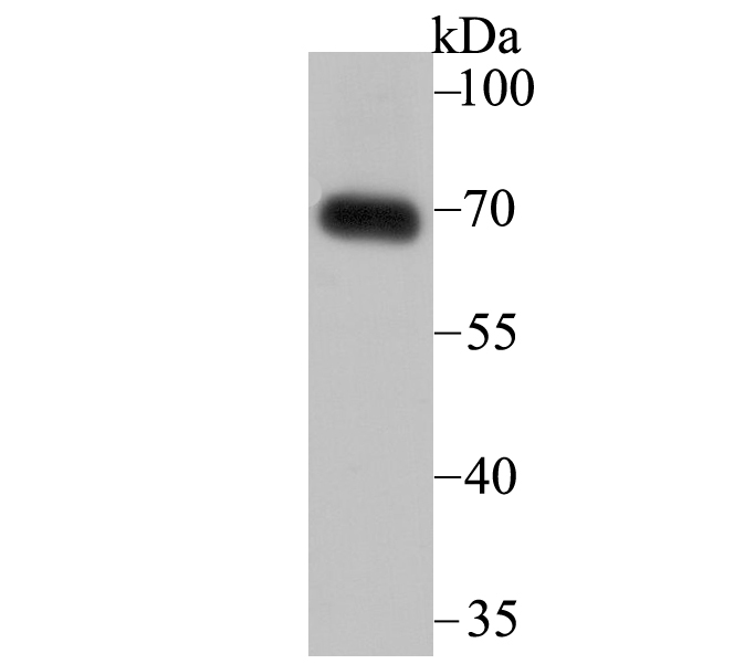
| Molekulargewicht: | Calculated MW 63 kDa |
| UniProt: | Q13596 |
| Puffer: | 1*TBS (pH7.4), 1% rAlbumin, 40% Glycerol, 0.05% Sodium Azide |
| Reinheit: | ProA affinity purified. |
| Formulierung: | Liquid |
| Application Verdünnung: | WB: 1:1,000-1:2,000 FC: 1:50-1:100 ICC/IF: 1:50-1:200 IHC: 1:50-1:200 |
|
|
Western blot analysis of SNX1 on human skin tissue lysate using anti-SNX1 antibody at 1/2000 dilution |
|
|
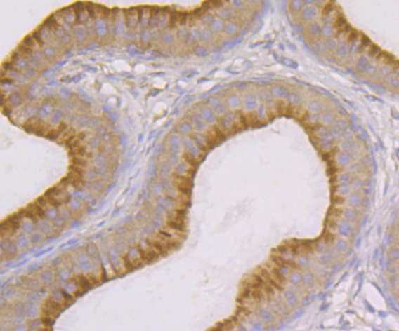
Immunohistochemical analysis of paraffin-embedded rat epididymis tissue using anti-SNX1 antibody. Counter stained with hematoxylin |
|
|
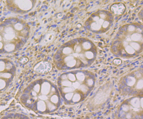
Immunohistochemical analysis of paraffin-embedded human colon tissue using anti-SNX1 antibody. Counter stained with hematoxylin |
|
|
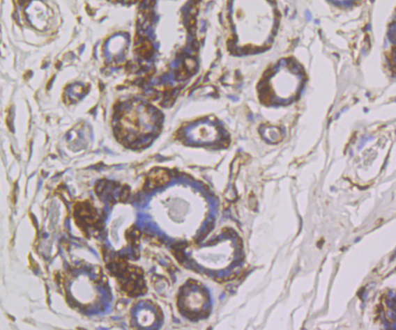
Immunohistochemical analysis of paraffin-embedded human breast cancer tissue using anti-SNX1 antibody. Counter stained with hematoxylin |
|
|
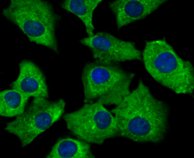
ICC staining SNX1 in A549 cells (green). The nuclear counter stain is DAPI (blue). Cells were fixed in paraformaldehyde, permeabilised with 0.25% Triton X100/PBS |
|
|
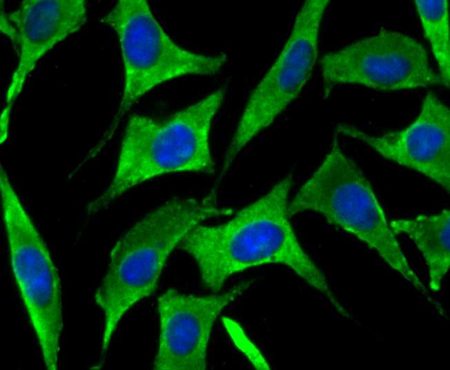
ICC staining SNX1 in SH-SY-5Y cells (green). The nuclear counter stain is DAPI (blue). Cells were fixed in paraformaldehyde, permeabilised with 0.25% Triton X100/PBS |
|
|
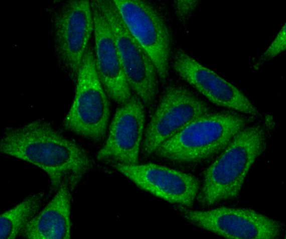
ICC staining SNX1 in SiHa cells (green). The nuclear counter stain is DAPI (blue). Cells were fixed in paraformaldehyde, permeabilised with 0.25% Triton X100/PBS |
|
|
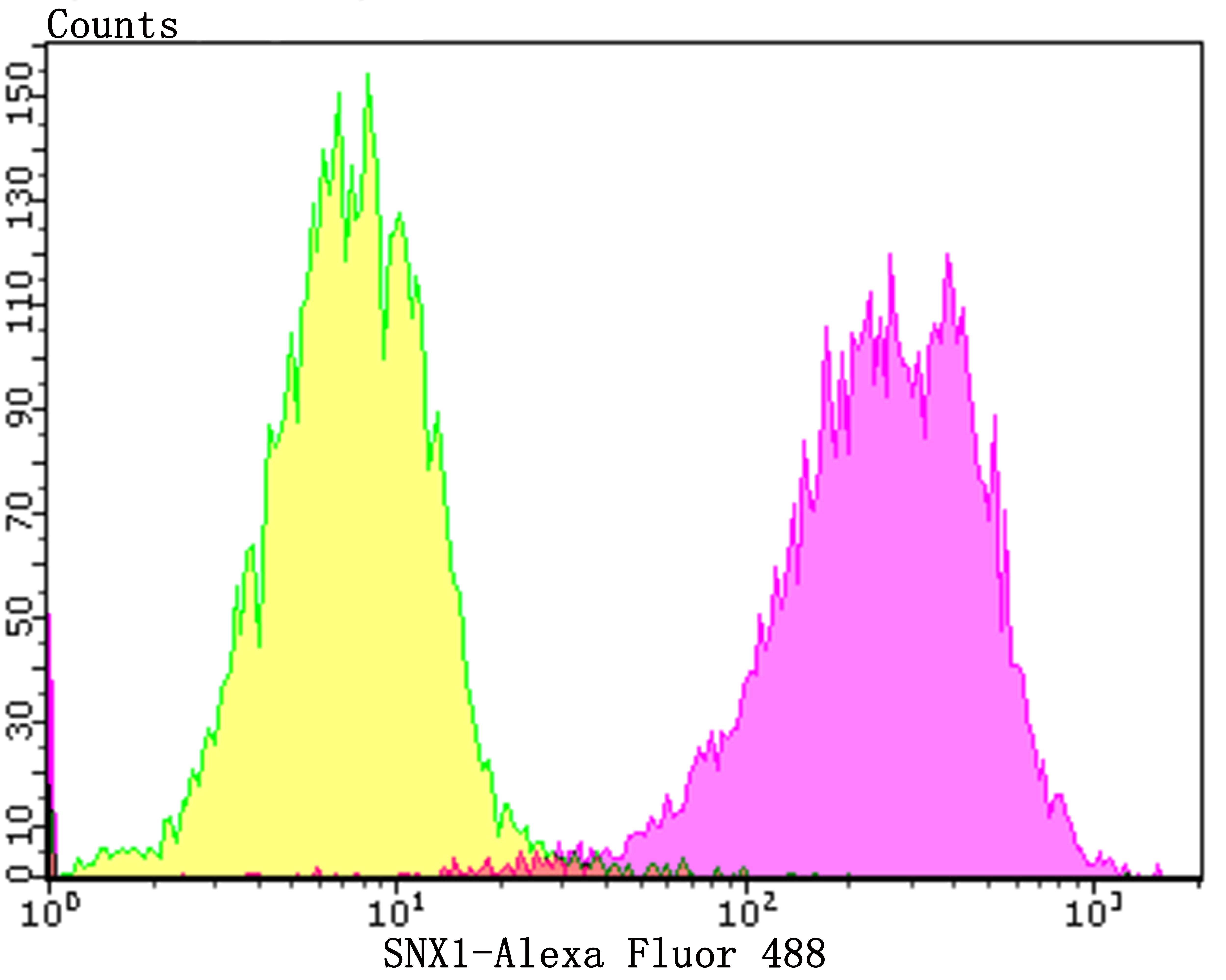
Flow cytometric analysis of SiHa cells with SNX1 antibody at 1/100 dilution (purple) compared with an unlabelled control (cells without incubation with primary antibody, yellow). Alexa Fluor 488-conjugated goat anti-rabbit IgG was used as the secondary antibody |
Produktgarantie und fachkundiger Support
