![]()
|
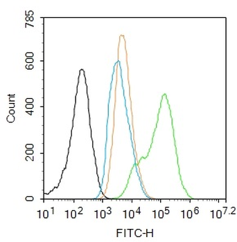
Blank control: A431. Primary Antibody (green line): Rabbit Anti-phospho-NFKB p65 (Thr435) antibody (orb6506), dilution: 1 µg/10 6 cells, Isotype Control Antibody (orange line): Rabbit IgG. Secondary Antibody: Goat anti-rabbit IgG-FITC, dilution: 1 µg/Test. Protocol, The cells were fixed with 4% PFA (10 min at room temperature) and then permeabilized with 90% ice-cold methanol for 20 min at -20C. The cells were then incubated in 5% BSA to block non-specific protein-protein interactions for 30 min at room temperature. Cells stained with Primary Antibody for 30 min at room temperature. The secondary antibody used for 40 min at room temperature. Acquisition of 20000 events was performed. |
![]()
|
Blank control: MCF7. Primary Antibody (green line): Rabbit Anti-phospho-NFKB p65 (Thr435) antibody (orb6506), dilution: 2 µg/10 6 cells, Isotype Control Antibody (orange line): Rabbit IgG. Secondary Antibody: Goat anti-rabbit IgG-AF647, dilution: 1 µg/Test. Protocol, The cells were fixed with 4% PFA (10 min at room temperature) and then permeabilized with 90% ice-cold methanol for 20 min at -20C. The cells were then incubated in 5% BSA to block non-specific protein-protein interactions for 30 min at room temperature. Cells stained with Primary Antibody for 30 min at room temperature. The secondary antibody used for 40 min at room temperature. Acquisition of 20000 events was performed. |
![]()
|
Blank control: Mouse spleen. Primary Antibody (green line): Rabbit Anti-phospho-NFKB p65 (Thr435) antibody (orb6506), dilution: 2 µg/10 6 cells, Isotype Control Antibody (orange line): Rabbit IgG. Secondary Antibody: Goat anti-rabbit IgG-AF488, dilution: 1 µg/Test. Protocol, The cells were fixed with 4% PFA (10 min at room temperature) and then permeabilized with 90% ice-cold methanol for 20 min at -20C. The cells were then incubated in 5% BSA to block non-specific protein-protein interactions for 30 min at room temperature. Cells stained with Primary Antibody for 30 min at room temperature. The secondary antibody used for 40 min at room temperature. Acquisition of 20000 events was performed. |
![]()
|
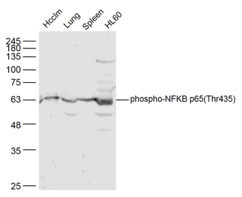
Sample: Hcclm3 Cell (Human) Lysate at 40 ug, Lung (Mouse) Lysate at 40 ug, Spleen (Mouse) Lysate at 40 ug, HL60 Cell (Human) Lysate at 40 ug, Primary: Anti-phospho-NFKB p65 (Thr435) (orb6506) at 1/300 dilution, Secondary: IRDye800CW Goat Anti-Rabbit IgG at 1/20000 dilution, Predicted band size: 61 kD, Observed band size: 61 kD. |
![]()
|
Tissue/Cell: Hela cell, 4% Paraformaldehyde-fixed, Triton X-100 at room temperature for 20 min, Blocking buffer (normal goat serum) at 37C for 20 min, Antibody incubation with (phospho-NFKB p65 (Thr435)) polyclonal Antibody, Unconjugated (orb6506) 1:100, 90 minutes at 37C, followed by a FITC conjugated Goat Anti-Rabbit IgG antibody at 37C for 90 minutes, DAPI (blue) was used to stain the cell nuclei. |
![]()
|
Tissue/Cell: MCF7 cell, 4% Paraformaldehyde-fixed, Triton X-100 at room temperature for 20 min, Blocking buffer (normal goat serum) at 37C for 20 min, Antibody incubation with (phospho-NFKB p65 (Thr435)) polyclonal Antibody, Unconjugated (orb6506) 1:100, 90 minutes at 37C, followed by a FITC conjugated Goat Anti-Rabbit IgG antibody at 37C for 90 minutes, DAPI (blue) was used to stain the cell nuclei. |