Mouse monoclonal antibody supplied in crude ascites with 0.09% (W/V) sodium azide.
Target-Kategorie:
SDC1
Application Verdünnung:
WB: 1:500-8000
Confocal immunofluorescent analysis of CD138 Antibody (C-term) (Ascites) with U266 cell followed by Alexa Fluor 488-conjugated goat anti-mouse lgG (green). DAPI was used to stain the cell nuclear (blue).
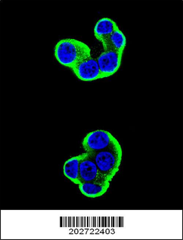
Confocal immunofluorescent analysis of CD138 Antibody (C-term) (Ascites) with HepG2 cell followed by Alexa Fluor 488-conjugated goat anti-mouse lgG (green).DAPI was used to stain the cell nuclear (blue).
Confocal immunofluorescent analysis of CD138 Antibody (C-term) (Ascites) with T47D cell followed by Alexa Fluor 488-conjugated goat anti-mouse lgG (green).DAPI was used to stain the cell nuclear (blue).
CD138 Antibody (C-term) (Ascites) western blot analysis in HepG2 cell line lysates (35 µg/lane). This demonstrates the CD138 antibody detected the CD138 protein (arrow).
CD138 Antibody (C-term) (Ascites) flow cytometric analysis of U266 cells (right histogram) compared to a negative control cell (left histogram).Alexa Fluor 488-conjugated donkey anti-mouse lgG secondary antibodies were used for the analysis
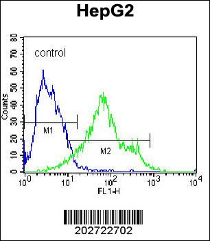
CD138 Antibody (C-term) (Ascites) flow cytometric analysis of HepG2 cells (right histogram) compared to a negative control cell (left histogram).Alexa Fluor 488-conjugated donkey anti-mouse lgG secondary antibodies were used for the analysis
CD138 Antibody (C-term) (Ascites) flow cytometric analysis of T47D cells (right histogram) compared to a negative control cell (left histogram).Alexa Fluor 488-conjugated donkey anti-mouse lgG secondary antibodies were used for the analysis
* Mehrwertsteuer und Versandkosten nicht enthalten. Irrtümer und Preisänderungen vorbehalten